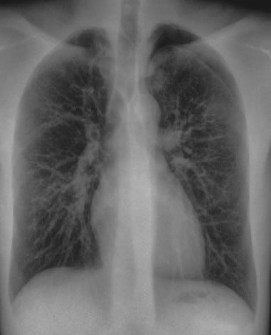
Chest radiography is one of the most widely used imaging examinations in medicine. It plays an important role in evaluating lung conditions, monitoring disease progression, and assessing thoracic structures.
However, overlapping anatomical structures such as ribs and clavicles may obscure clinically relevant findings in conventional chest radiographs.
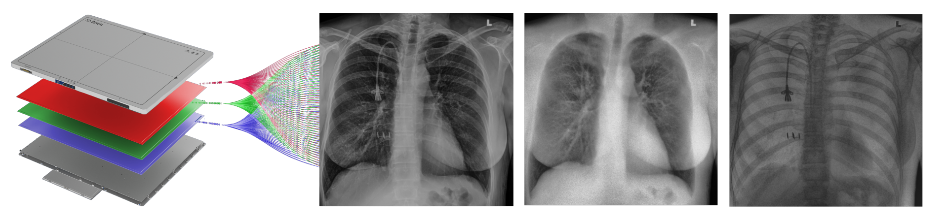
Spectral X-ray imaging expands the information available in routine chest radiography. KA Imaging’s Reveal™ platform uses SpectralDR® technology to generate multiple image types from a single exposure, including conventional, bone, and soft-tissue images, while maintaining the same workflow and radiation exposure as standard radiography.
Solutions for Chest Imaging
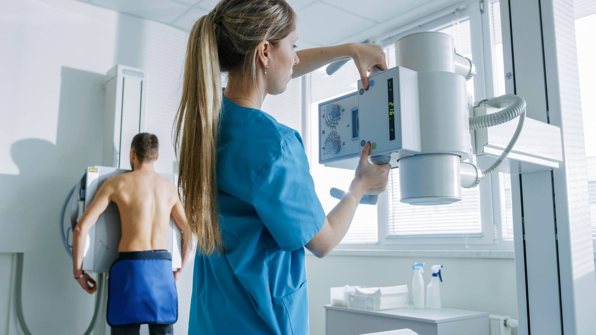
Reveal™ spectral imaging solutions can be used in emergency departments and trauma centers.
Chest radiography is used to evaluate a wide range of pulmonary and thoracic conditions.
evaluation of lung opacities
assessment of pneumothorax
monitoring pneumonia or infection
evaluation of lung nodules or lesions
Spectral imaging can enhance visualization of lung structures by separating bone and soft-tissue information into multiple images generated from a single exposure. Clinical studies using the Reveal 35C suggest:
33%
more pneumonia cases found compared to X-ray thanks to dual-energy images⁴
43%
more lesion visibility when dual-energy images were included³
This additional imaging information may help clinicians interpret chest radiographs more clearly.
In this study, the conventional DR was read as normal. Upon viewing the Dual-Energy Soft Tissue image, the radiologist noticed a highlighted focal opacity indicative of pneumonia (confirmed on CT).
Trusted by Users
Patrik RogallaRadiologist
In the first 34 patients, Reveal™’s dual energy capabilities have already helped find pneumonia with higher confidence. We are continuing to image and will be adding a second system to increase our imaging capacity.
Vikram VenkateshRadiologist
Dual-energy images from the Reveal™ detector helped find lung nodules and scarring that were hard to pick up on the X-ray image. The dual-energy images also showed positioning of lines and tubes very clearly, which can be difficult to appreciate on portable X-ray images.
Spectral Imaging for Chest Imaging
Reveal™ 35C detectors use SpectralDR® technology to acquire multiple spectral images from a single X-ray exposure.
Each exposure produces:
a conventional digital radiography image
a bone image
a soft-tissue image
These images allow clinicians to separate overlapping anatomical structures and visualize areas that may be less visible in conventional radiographs.
Unlike traditional dual-energy imaging techniques that require multiple exposures, SpectralDR captures these images simultaneously with a single exposure, eliminating motion artifacts and maintaining the same clinical workflow as standard portable radiography.
Key Clinical Scenarios in Chest Imaging
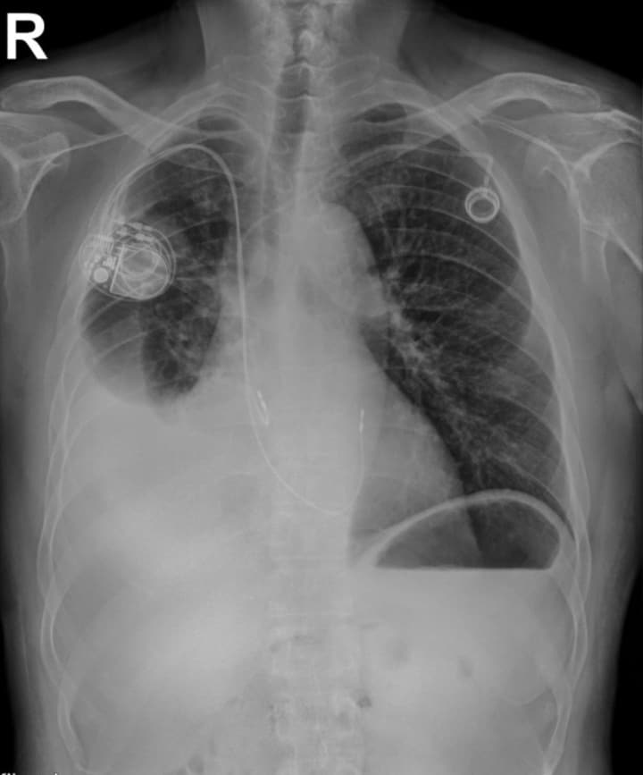
Lung Lesion Visibility
Soft-tissue spectral images may improve visualization of pulmonary nodules and lesions that overlap with rib structures.
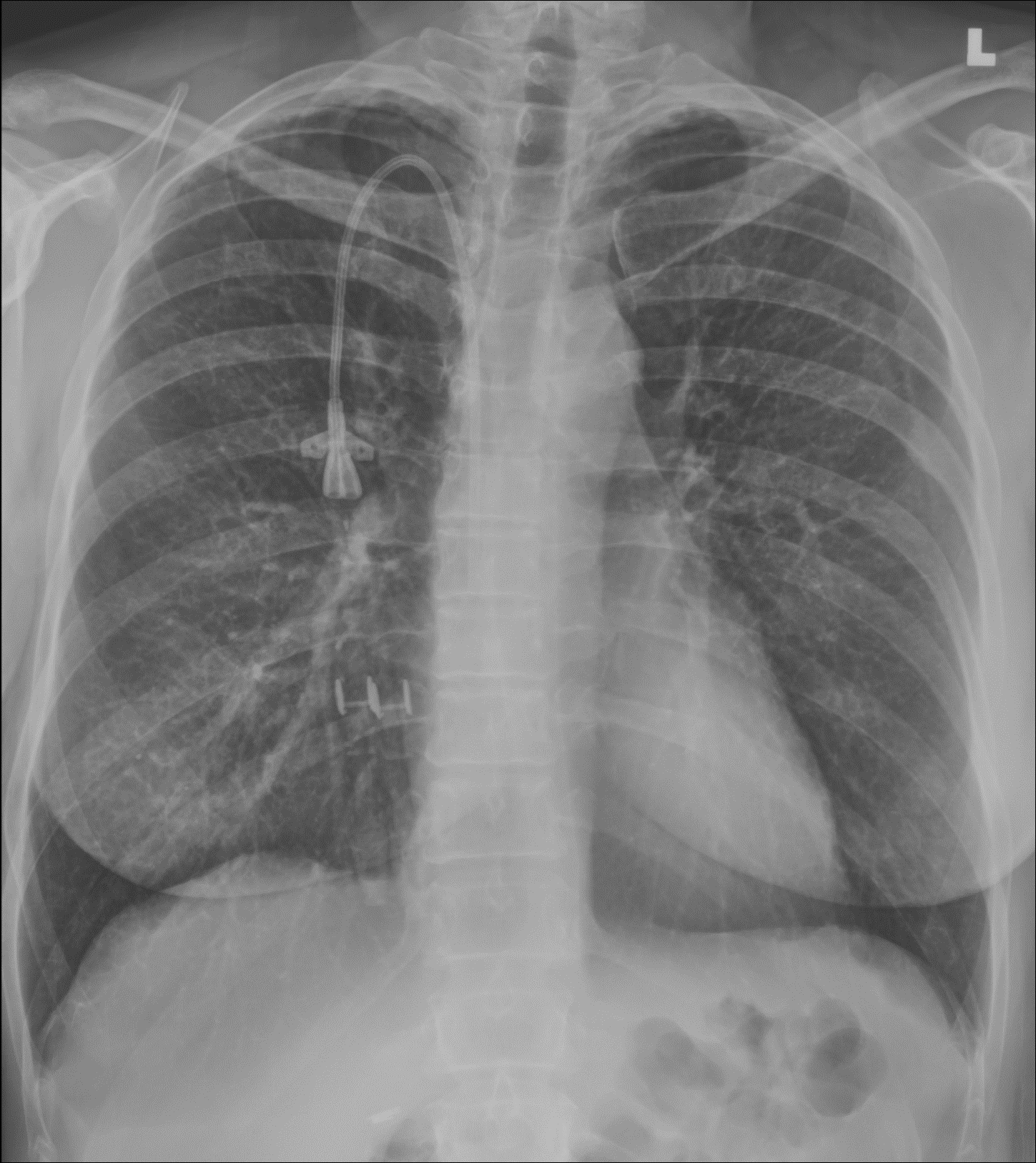
In this study, the dual energy soft tissue image revealed a suspicious soft tissue mass. In this case, the mass was overlapping the in-dwelling device (pacemaker).
Pneumonia Evaluation
Spectral imaging can enhance visualization of lung opacities by separating bone structures from soft tissue.
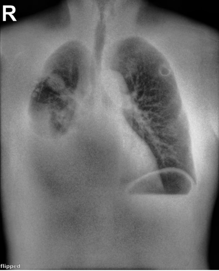
Pneumothorax Visualization
Soft-tissue images reduce rib overlap and may improve visualization of pneumothorax in chest radiographs.
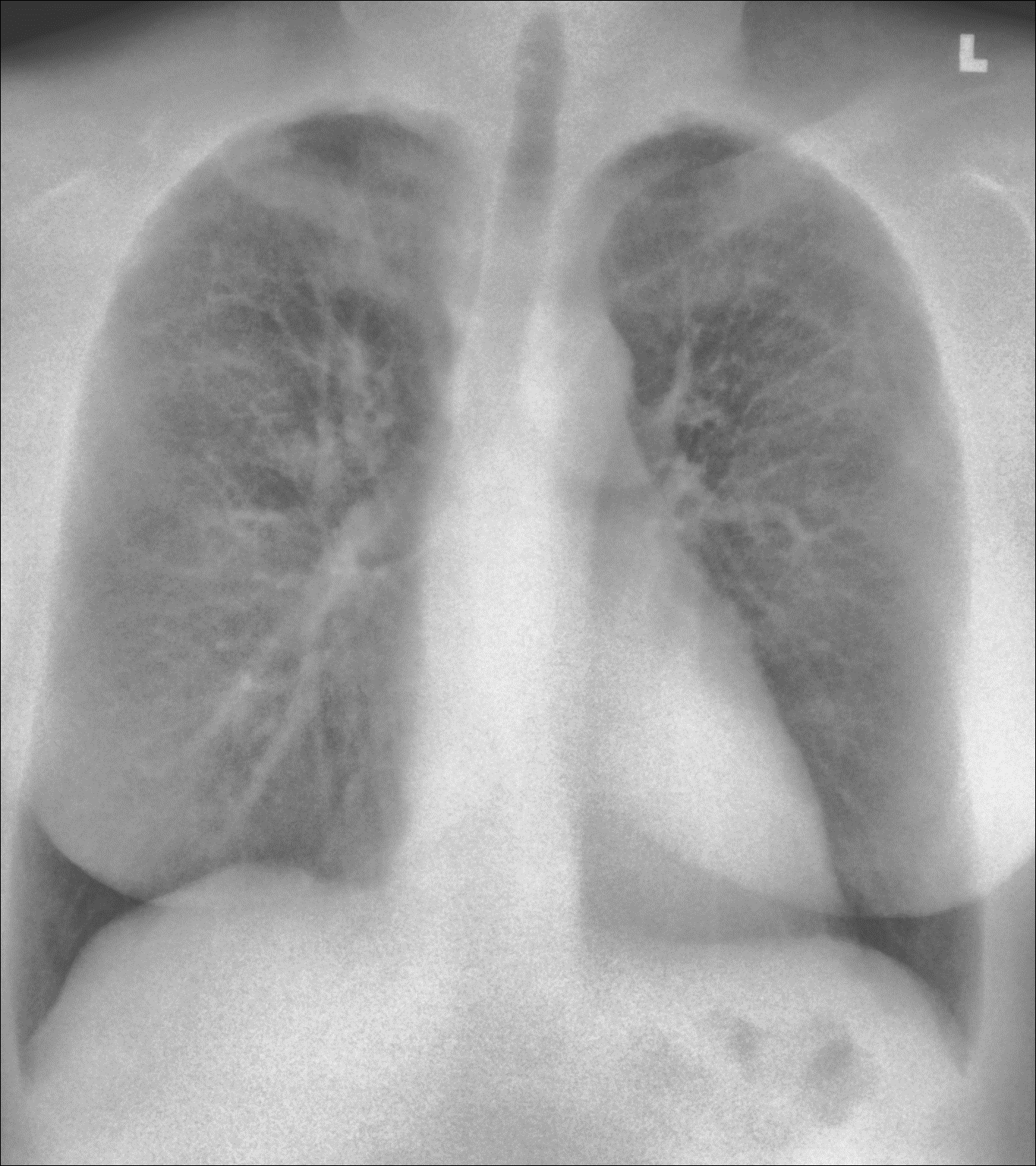
This 26-year-old male presented to the ER at 3am, with difficulty breathing and chest pain. The Reveal 35C Spectral DR detector was used to generate all 3 images. A left apical pneumothorax can be visualized, and is best seen in the Soft Tissue image.