
SpectralDR for Global Health Imaging
Expanding Imaging Capabilities Where Advanced Modalities Are Limited
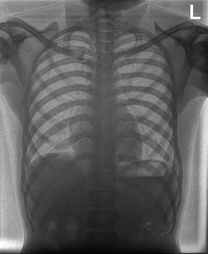
Chest X-ray is often the primary imaging modality in healthcare environments where access to CT and other advanced technologies is limited.
Clinical Context in Global Health
X-ray imaging is commonly used in many regions for evaluating:
- respiratory diseases
- thoracic abnormalities
- musculoskeletal injuries
- monitoring disease progression
Expanding the information available from routine X-ray exams may help clinicians make more informed decisions while working within limited healthcare resources.
Clinical Case
Solutions for Global Health Imaging
These systems can be deployed in a wide range of environments, including hospitals, regional clinics, and mobile healthcare units.
Relevant solutions include:
Spectral Imaging at the Point of Care
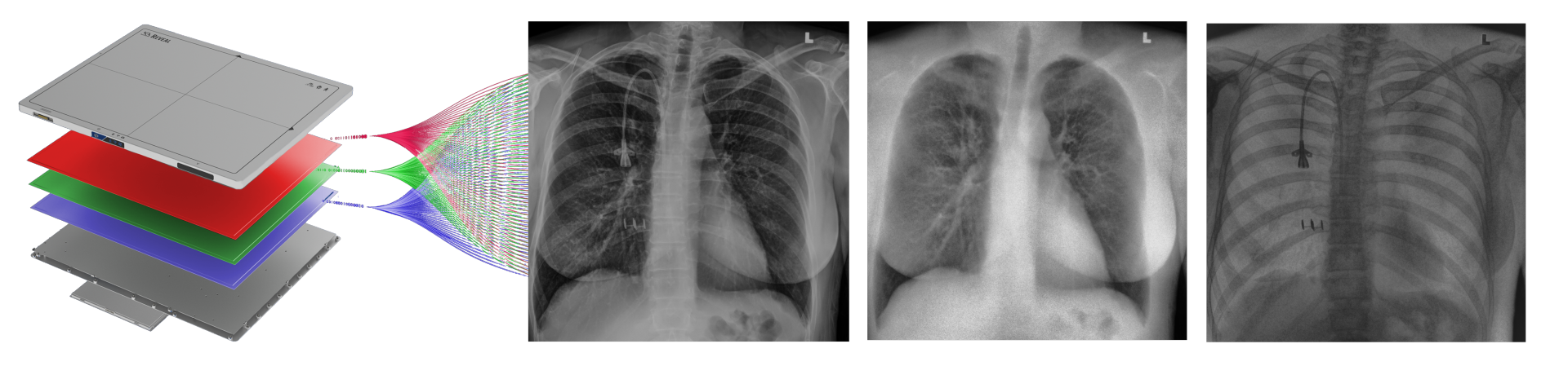
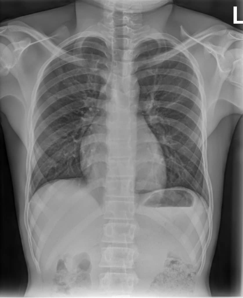
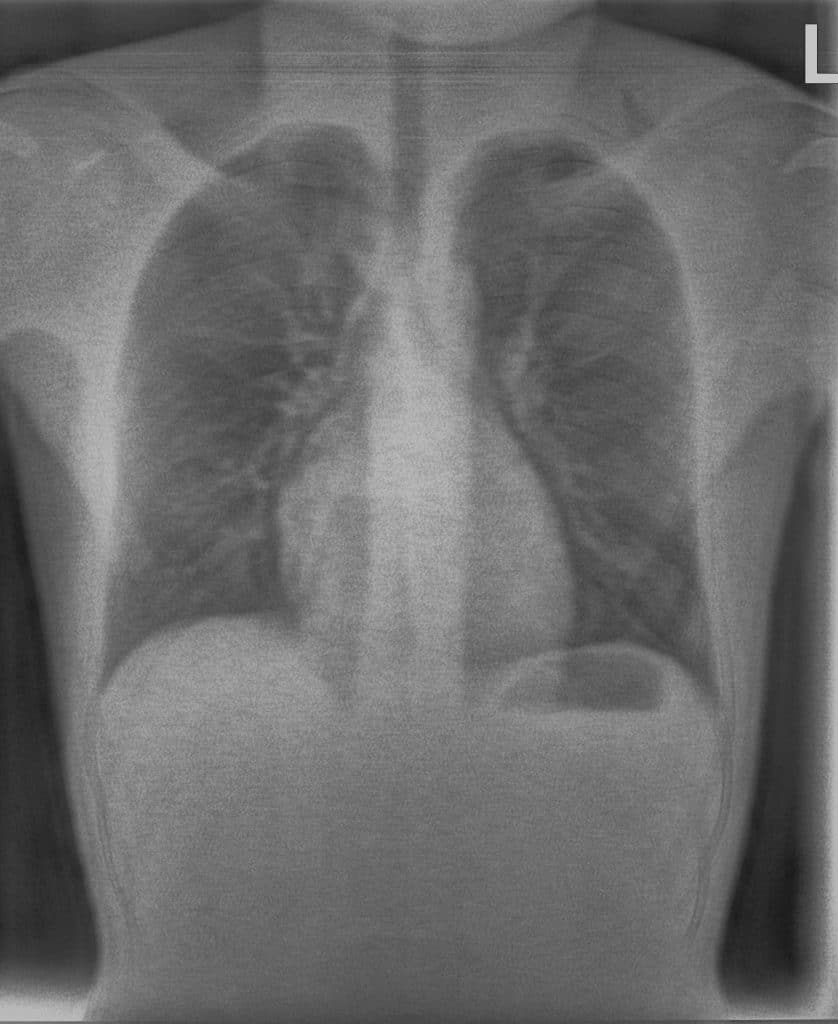
Reveal™ detectors generate three images from a single X-ray exposure:
- conventional radiograph
- bone image
- soft-tissue image
These images allow clinicians to examine anatomical structures from different perspectives and may provide additional insight when evaluating radiographs in environments where other imaging modalities are not available.
Because these images are generated during a routine radiography exam, spectral imaging can be incorporated into existing imaging workflows.
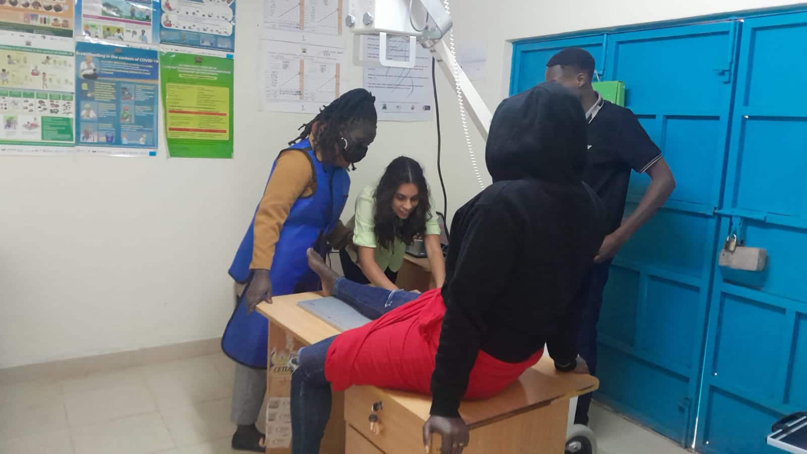
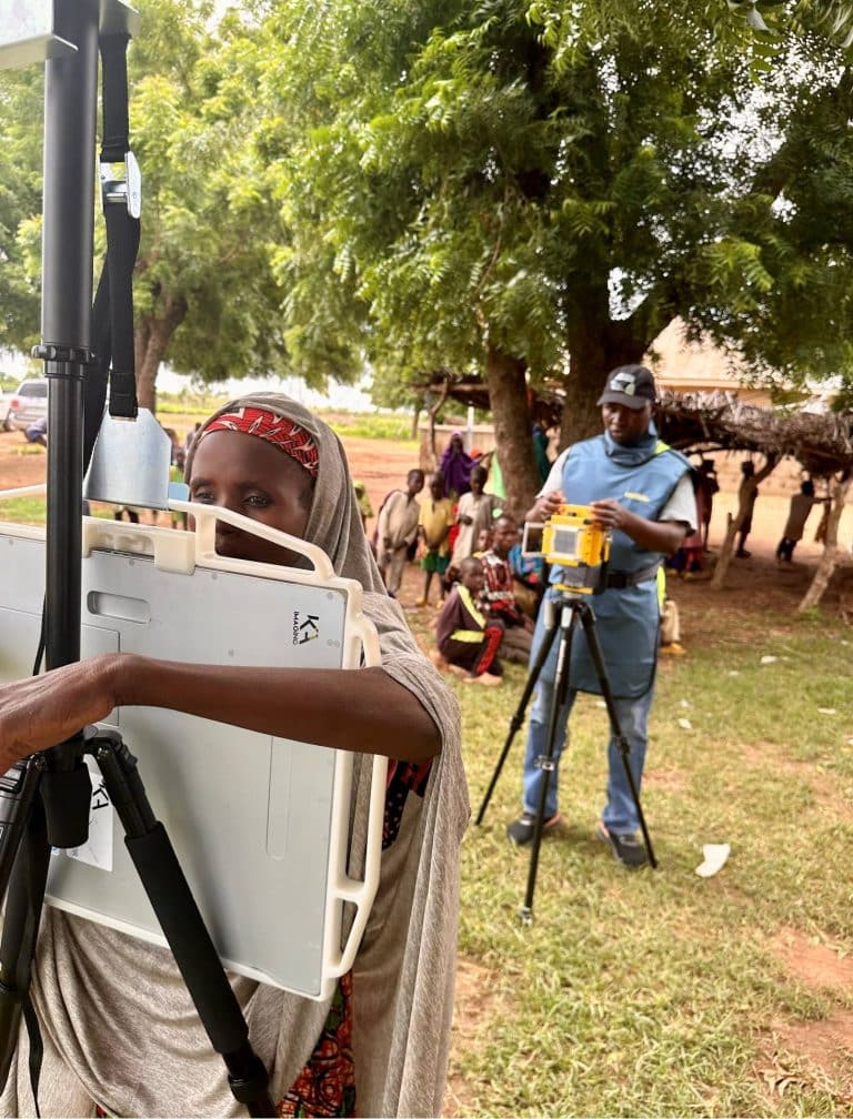
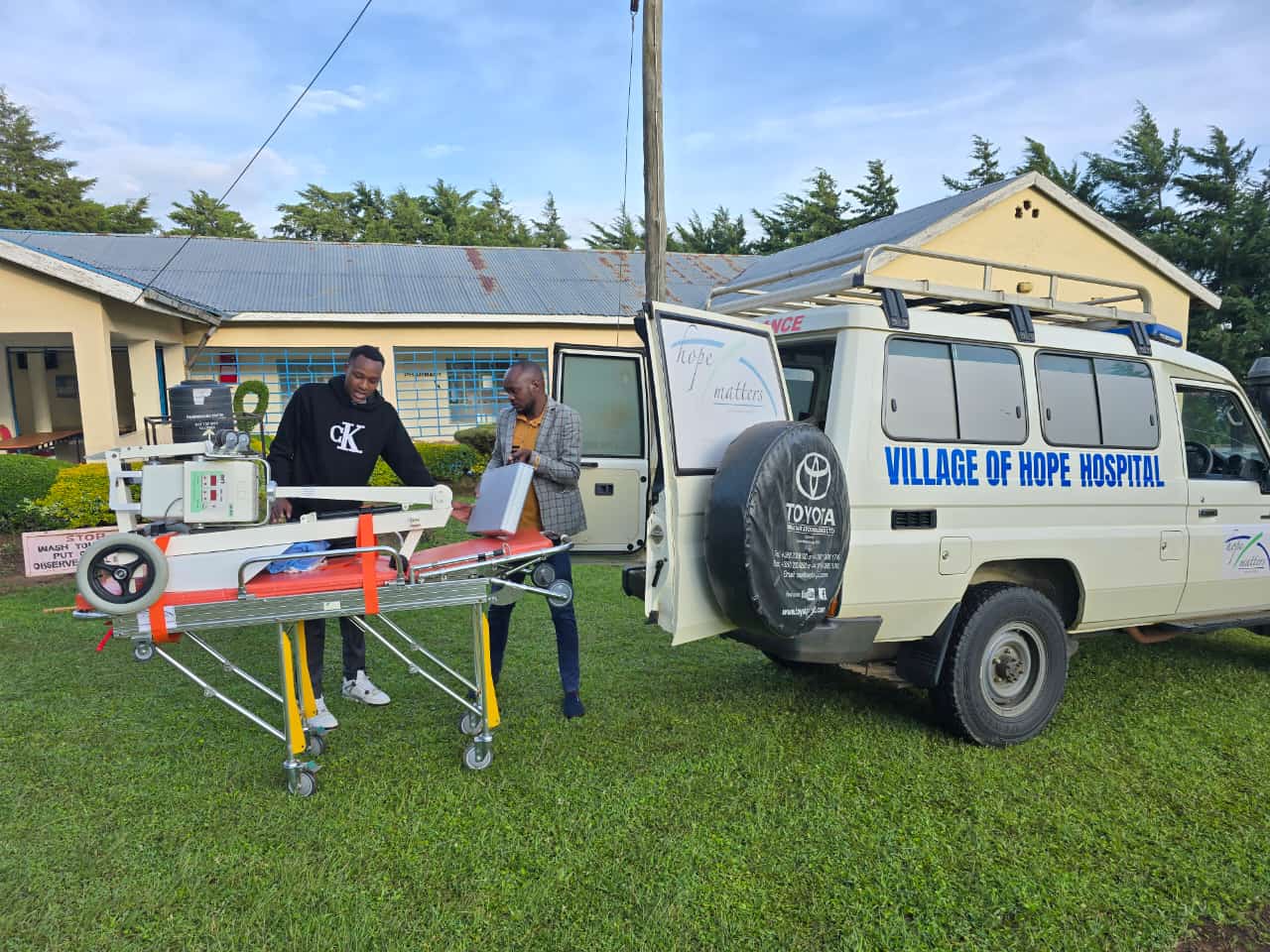
Case Study: bringing advanced imaging to Kenya
KA Imaging has partnered with the Kenyan Kids Foundation Canada to bring advanced X-ray imaging technology to a remote area of Kenya. In August 2024, our team traveled to install the Reveal 35C detector and an X-ray source, enabling access to dual-energy X-ray imaging in a region with limited medical resources. Watch to see how medical imaging technology can improve care in underserved communities.
Trusted by Users
Related Applications
Critical Care & Bedside Imaging
Chest Imaging
Emergency & Trauma Imaging

Expanding Access to Imaging Insight
Latest blogs
WATERLOO, Ontario, 20 July 2026 – KA Imaging has expanded its international footprint with regulatory…
Why Lateral Views Matter Lateral chest X-rays play an important role in diagnostic imaging. While…
Modern airport and cargo security systems are designed for throughput. Large-scale X-ray scanners inspect high…