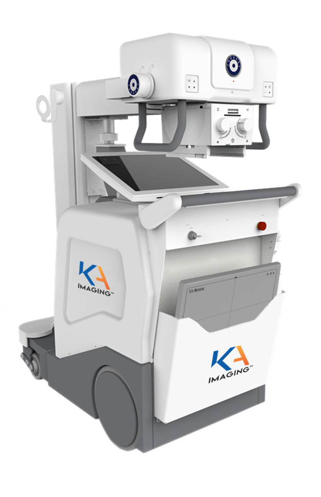
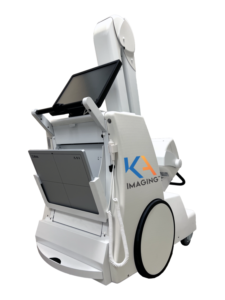
Reveal™ Portable Package 35C-Dragon
For timely and accurate bedside imaging. Our ultra portable solution with Reveal™ 35C andSpectralDR® technology.
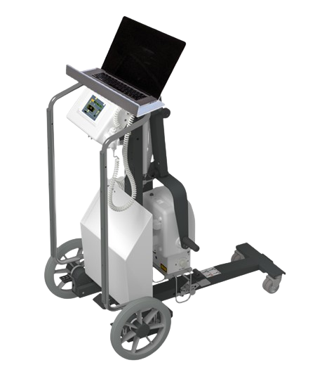
The Reveal™ Portable Package 35C-Dragon is an Analog Portable X-Ray Unit with Detector Retrofit. It combines the light weight of the Dragon X SPSL-HF-4.0 portable source with the exclusive single exposure dual-energy capabilities of KA Imaging’s Reveal™ 35C, powered by SpectralDR® technology. The package includes the Dragon portable source, the Reveal™ 35C detector with accessories and a laptop.
This affordable ultra-portable X-ray bundle goes beyond conventional imaging, providing clinicians with invaluable clinical insights for informed decision-making.
Timely and accurate bedside imaging: an ever-present need
Timely and accurate bedside imaging is essential for effective diagnosis and treatment. Sometimes, care is needed in areas that lack the traditional hospital infrastructure. Remote locations, disaster response, mass casualty incidents, you name it: the demand for accessible imaging solutions is ever-present.
Urban areas have their own challenges: critical care and emergency medicine, surgical procedures, long term homes and geriatric care, sports medicine and rehabilitation. The low mobility temporarily or permanently faced by patients requires high-quality imaging – preferably avoiding patient transport inside the hospital or other facilities.
The Reveal™ Portable Package 35C-Dragon is as a solution that can be easily deployed in a variety of environments, making it ideal for efficient healthcare delivery.
Product overview
Includes the Reveal™ 35C detector with SpectralDR® technology
Analog portable unit
Lightweight: 54kg
4 kW generator
Literature
SpectralDR® technology: supplemental subtracted images for better decision-making on the move

In addition to the DR, the SpectralDR® technology in the Reveal™ 35C detector offers quick access to supplemental bone and soft tissue images. No extra radiation is needed: all three images are created simultaneously with a single standard X-ray dose.
Get the most of your mobile X-ray system particularly in challenging environments like the ICU or the Emergency Room – where transporting patients carries clinical and operational hazards, increasing the chances of adverse events.
Dual-energy images are clinically proven1 to enhance the visualization of lung nodules, pneumonia, line and tube tips, pneumothorax, retained surgical objects and more.
Clinical Case
Discover a Hidden Focal Opacity in a Portable X-ray
This is a 51-year-old female leukemia (AML) patient. First, the conventional DR X-Ray was read as normal. After seeing the soft tissue image, the radiologist said they now noticed a highlighted focal opacity indicative of pneumonia (confirmed on CT). The diagnosis was confirmed positive for febrile neutropenic pneumonia.
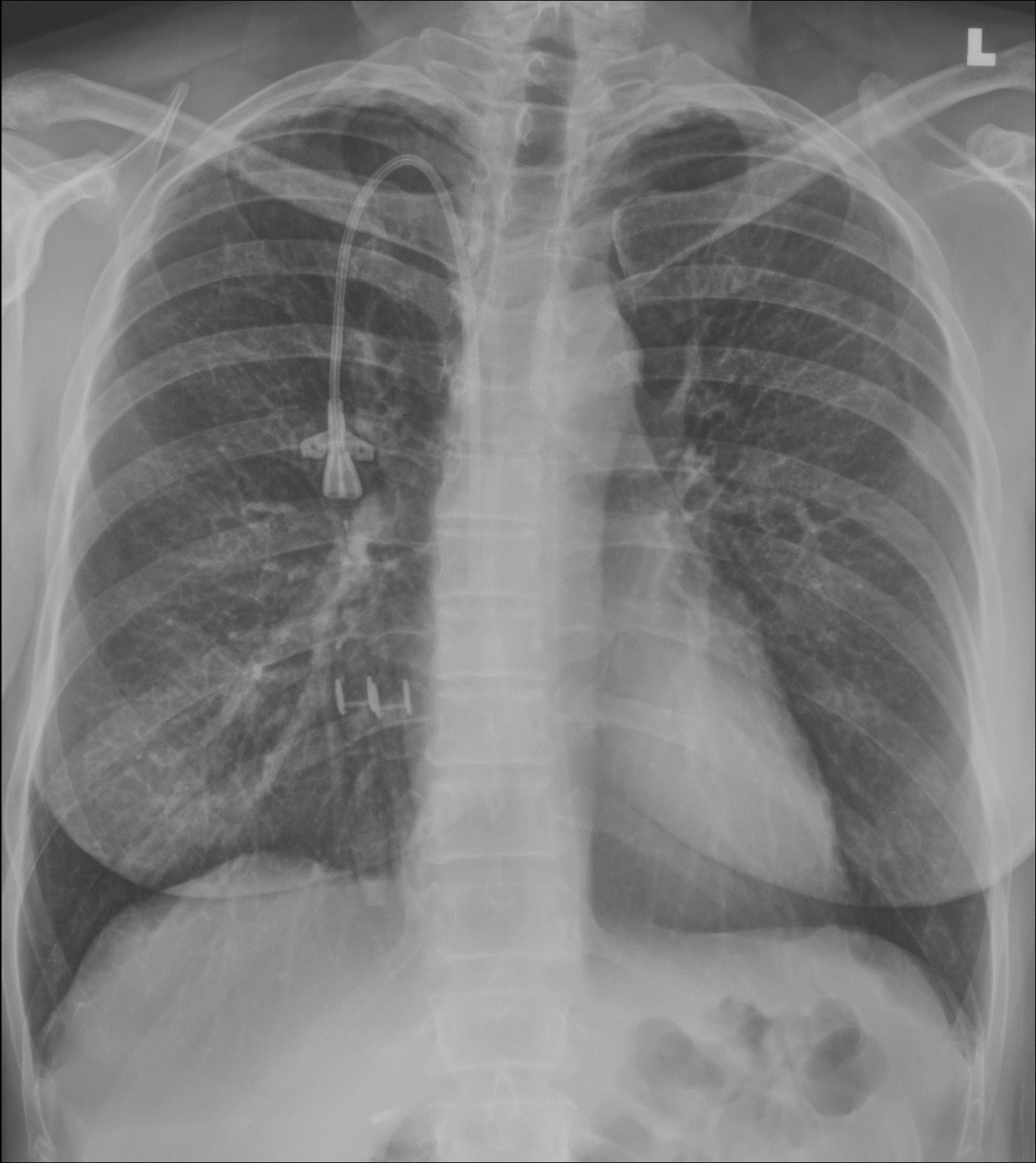
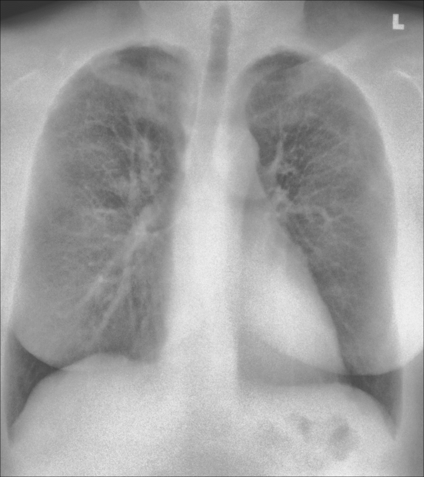
DR/SoftTissue Image
Clinical Case
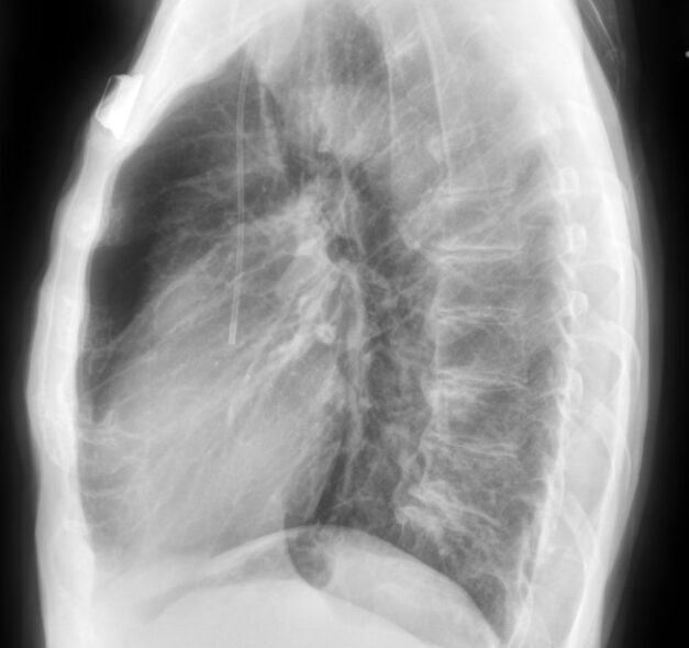
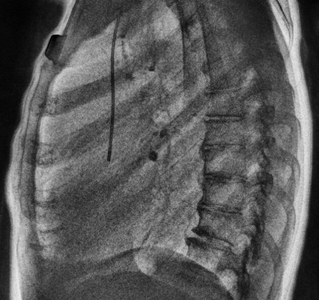
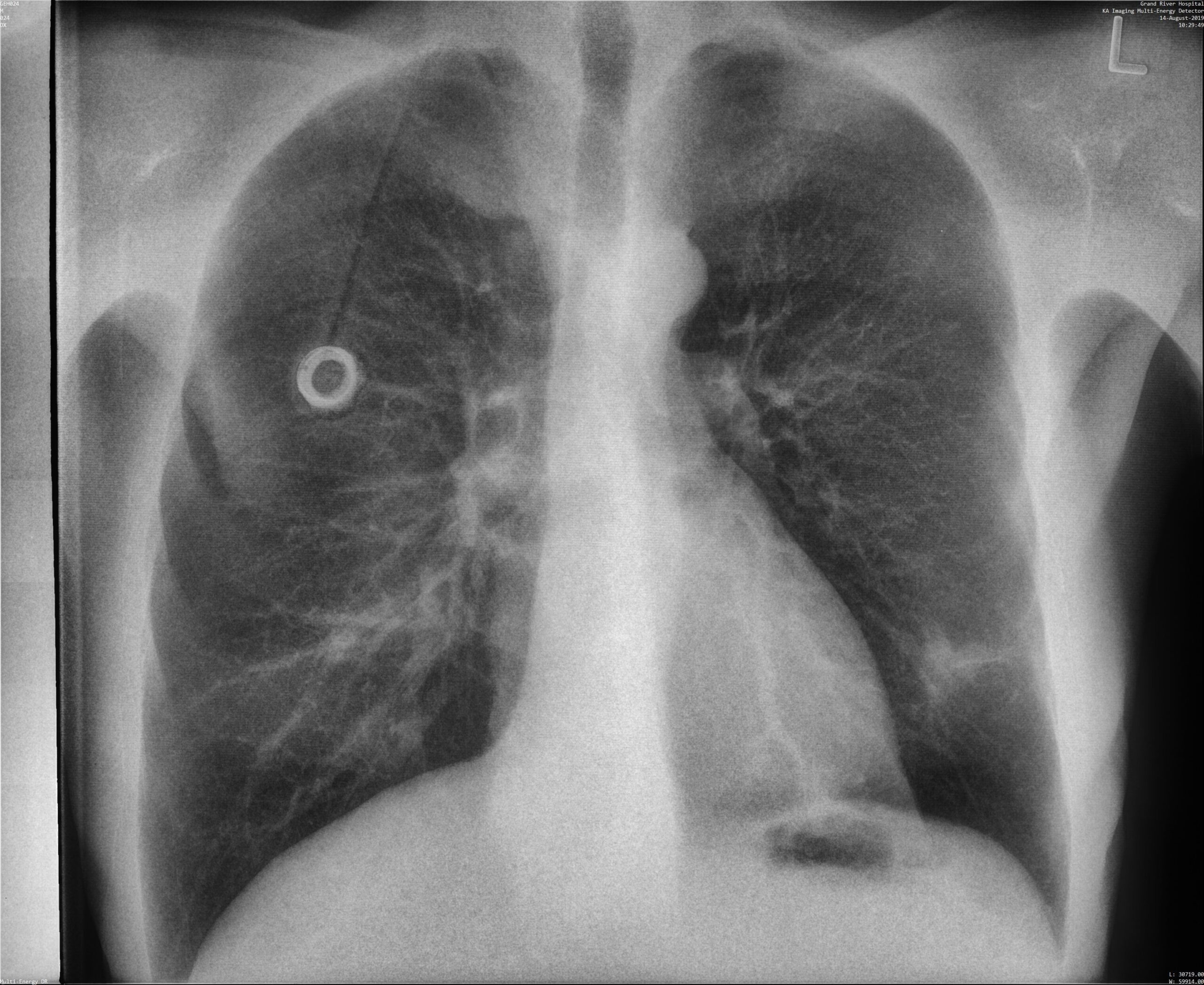
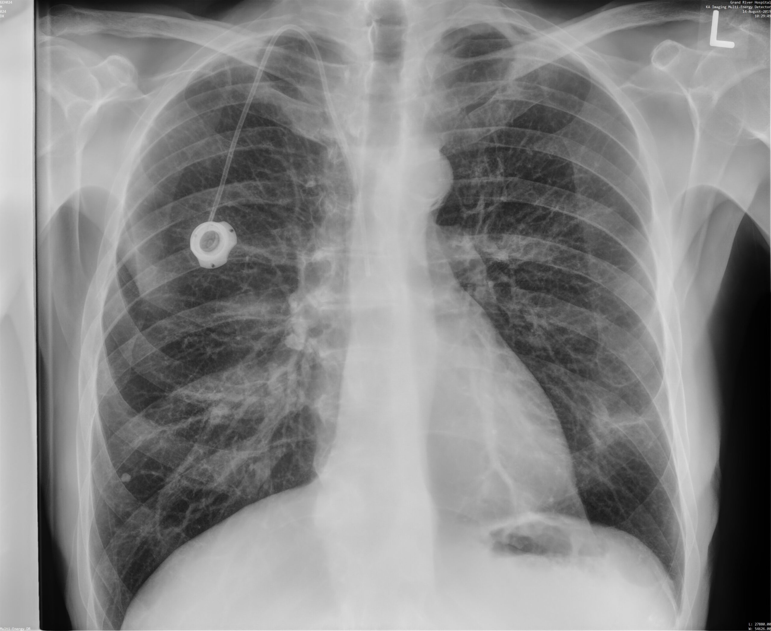
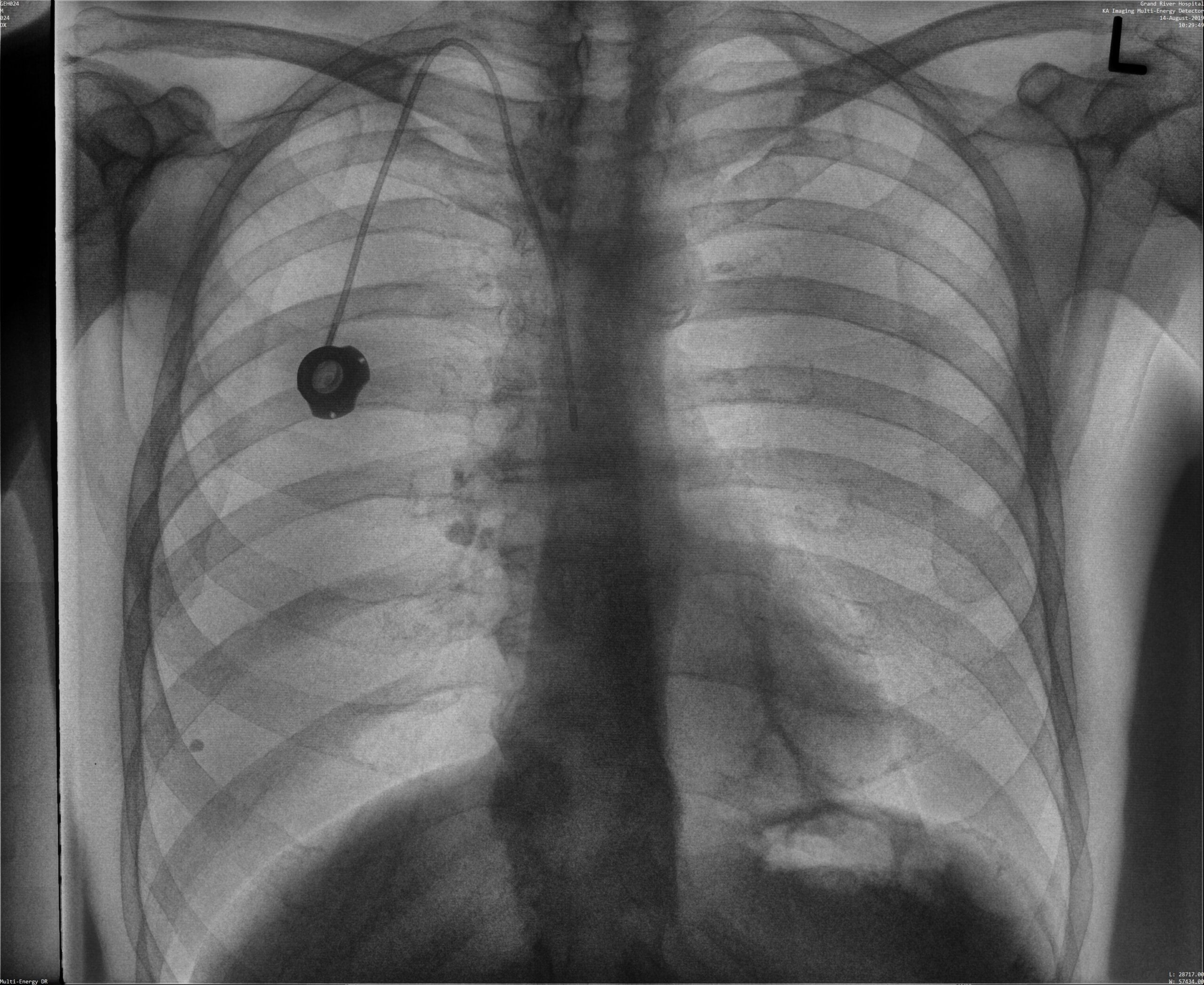
Better Line Delineation at the Bedside with no additional software
Portable imaging is taken to a new level with the ability to quickly and efficiently allow physicians to verify proper line and tube placements, at no extra dose to the patient, nor the need of add-on software. Take note of the high level of detail exhibited in the vascular wire mesh.
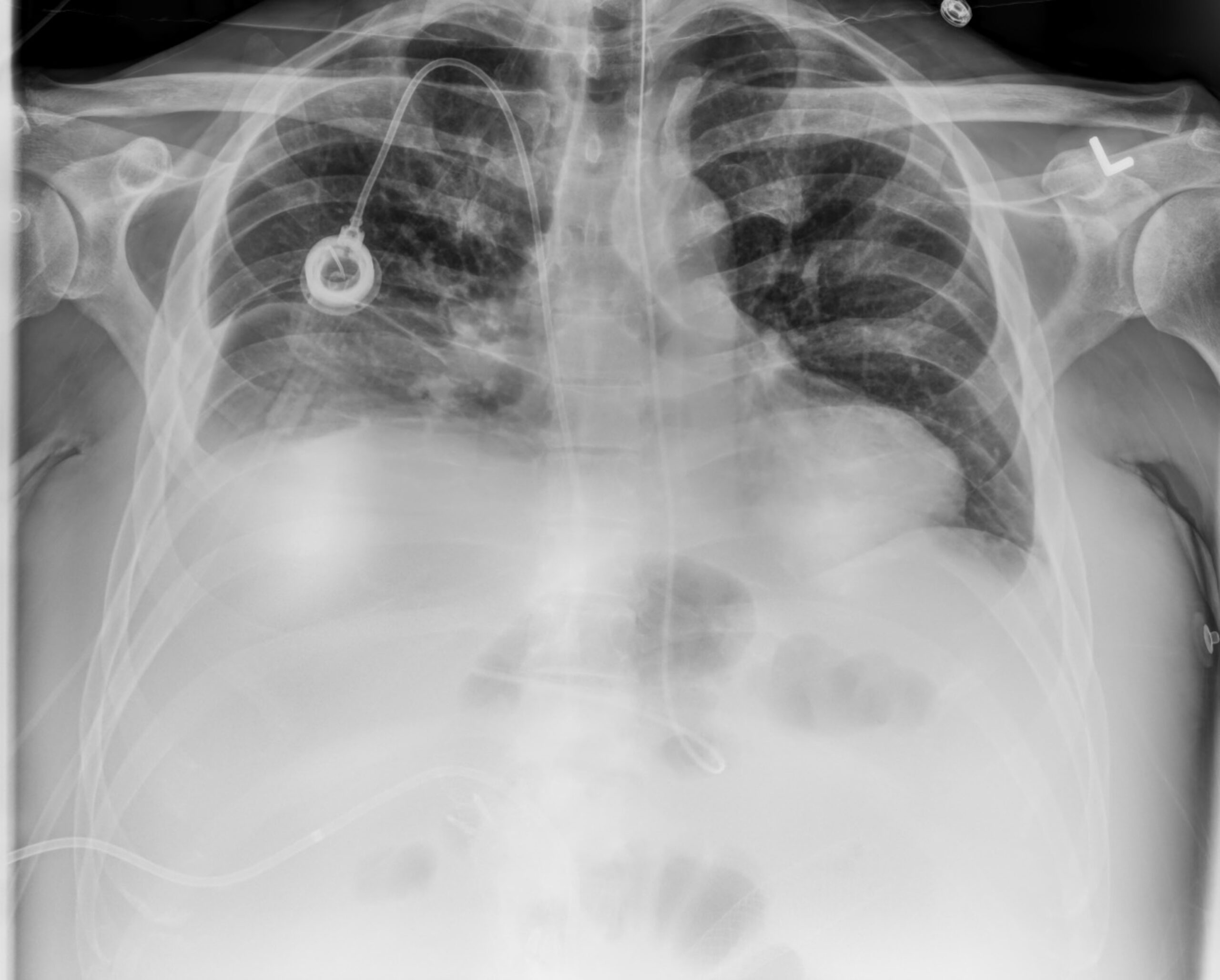
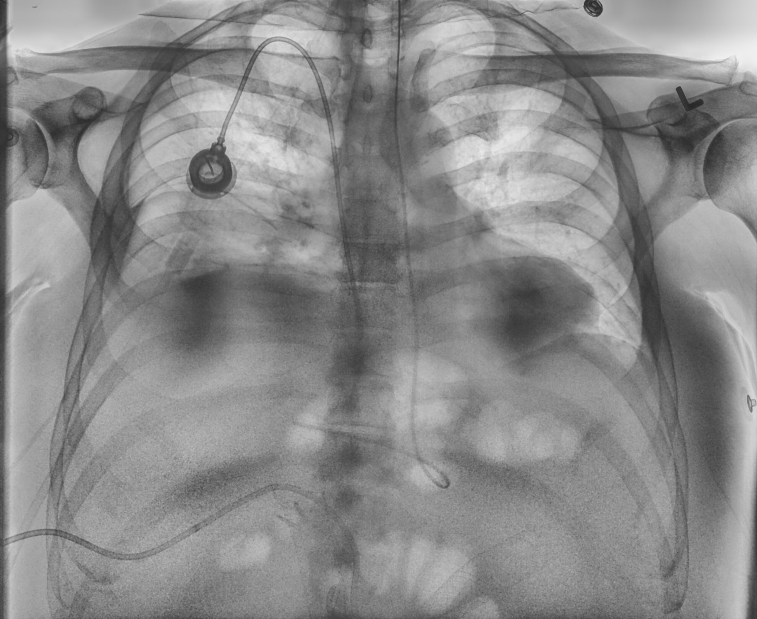
DR/Bone Image
Reveal™ 35C: proven results in diagnostic imaging
33% more pneumonia cases found
43% more lesion visibility
Better visibility of lines and tube tips⁵
Increased confidence in 57% of ICU cases
Increases revenue through incidental findings;
Success story
Learn How a Medium-Size Community Hospital Was Able to Reduce Follow-Up Imaging Thanks To SpectralDR® Technology.

Testimonials
Media Gallery



Latest blogs
Every budget season, hospitals ask imaging leaders to predict the future using yesterday’s tools. Capital…
Traditionally, X-ray technology has not been able to come to space, but the single exposure…
Dual-energy X-ray imaging has traditionally been associated with large, fixed radiography systems and complex workflows.…
The Dragon X SPSL-HF-4.0 portable source, as well as the Reveal™ 35C detector are both FDA cleared, Health Canada approved, and CE marked. Contact us for availability in your geographic area.
References:
Improved patient outcomes
1.1 (Lung Nodules) Oda, Seitaro, Kazuo Awai, Yoshinori Funama, Daisuke Utsunomiya, Yumi Yanaga, Koichi Kawanaka, Takeshi Nakaura et al. “Detection of small pulmonary nodules on chest radiographs: efficacy of dual-energy subtraction technique using flat-panel detector chest radiography.” Clinical radiology 65, no. 8 (2010): 609-615.
1.2 (Pneumothorax) Urbaneja, A., Dodin, G., Hoosu, G., et al. (2018) Added Value of Bone Subtraction in Dual-energy Digital Radiography in the Detection of Pneuomothorax: Impact of Reader Expertise and Medical Specialty. The Association of University Radiologists. Elsevier Inc.
1.3 (Pneumonia) Martini, Katharina, Marco Baessler, Stephan Baumueller, and Thomas Frauenfelder. “Diagnostic accuracy and added value of dual-energy subtraction radiography compared to standard conventional radiography using computed tomography as standard of reference.” PloS one 12, no. 3 (2017): e0174285.
1.4 (Tuberculosis) Sharma, Madhurima, Manavjit Singh Sandhu, Ujjwal Gorsi, Dheeraj Gupta, and Niranjan Khandelwal. “Role of digital tomosynthesis and dual energy subtraction digital radiography in detection of parenchymal lesions in active pulmonary tuberculosis.” European Journal of Radiology 84, no. 9 (2015): 1820-1827.
1.5 (Coronary Calcifications) Song, Yingnan, Hao Wu, Di Wen, Bo Zhu, Philipp Graner, Leslie Ciancibello, Haran Rajeswaran et al. “Detection of coronary calcifications with dual energy chest X-rays: clinical evaluation.” The International Journal of Cardiovascular Imaging (2020): 1-8.
- Maurino, S. L., Badano, A., Cunningham, I. A., & Karim, K. S. (2016, March). Theoretical and Monte Carlo optimization of a stacked three-layer flat-panel x-ray imager for applications in multi-spectral diagnostic medical imaging. In Medical Imaging 2016: Physics of Medical Imaging (Vol. 9783, p. 97833Z). International Society for Optics and Photonics.
- S. L. Maurino, K. S. Karim, V. Venkatesh. Diagnostic value of single-exposure dual-energy subtraction radiography in lung lesion detection: initial results. European Congress of Radiology-ECR 2022, 2022.
- Sanchez F, Kandel S, May M, Ronghe S, Rogalla P. Diagnostic value of dual-energy chest x-ray in immunocompromised patients to rule out pneumonia: initial results. European Congress of Radiology-ECR 2021, 2021.
- Rogalla P., Karim K. Added diagnostic value of portable dual-energy chest X-ray in a non-radiological reviewing environment. Radiological Society of North America-RSNA 2022, 2022
- Kuhlman, Janet E., Jannette Collins, Gregory N. Brooks, Donald R. Yandow, and Lynn S. Broderick. “Dual-energy subtraction chest radiography: what to look for beyond calcified nodules.” Radiographics 26, no. 1 (2006): 79-92.
- Manji, Farheen, Jiheng Wang, Geoff Norman, Zhou Wang, and David Koff. “Comparison of dual energy subtraction chest radiography and traditional chest X-rays in the detection of pulmonary nodules.” Quantitative imaging in medicine and surgery 6, no. 1 (2016): 1.
- Karim S Karim, “Single Exposure, Digital Dual-Energy Subtraction X-Ray Ushers in a New Era of Diagnostic X-Ray Imaging,” Radiology Management, Mar/Apr 2021.

