
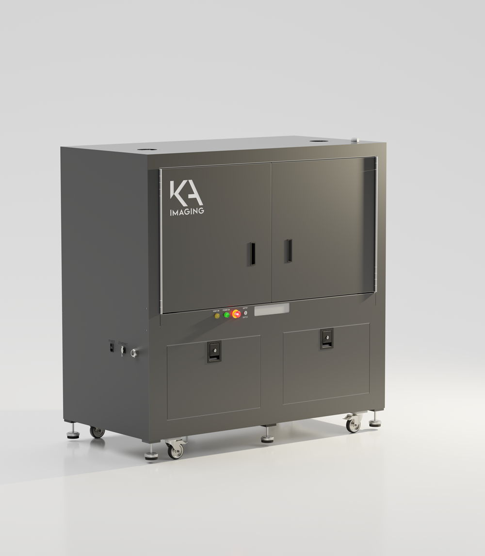
inCiTe™ 2.0 Configurable
3D X-ray Microscope
An innovative micro-CT system that integrates both phase contrast and spectral imaging capabilities.
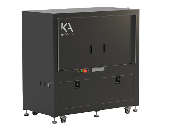
inCiTe™ 2.0: Configurable 3D X-ray Microscopy with Phase and Spectral Imaging
The inCiTe™ 2.0 3D X-ray Microscope extends phase-contrast imaging by integrating spectral imaging capabilities, enabling multi-material analysis within a single scan.
By combining phase and spectral information, inCiTe 2.0 supports improved differentiation of materials with similar densities, making it well-suited for complex structures and multi-material systems in industrial and research applications.
Designed for non-destructive testing and advanced analysis workflows, the system provides high-resolution 3D imaging while enabling deeper insight into material composition and structure.
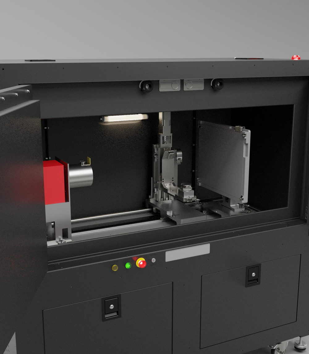
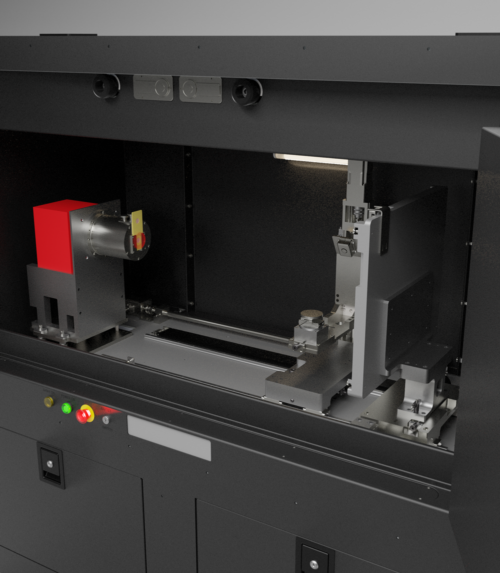
Product overview
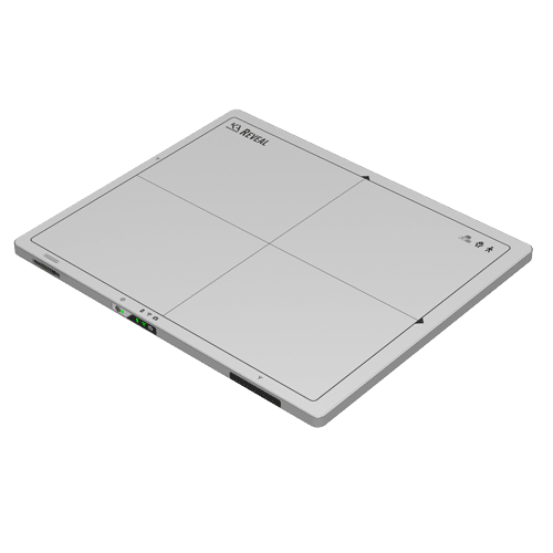
2 X-ray detector options: BrillianSe Hybrid Pixel Detector or Reveal Flat Panel Detector
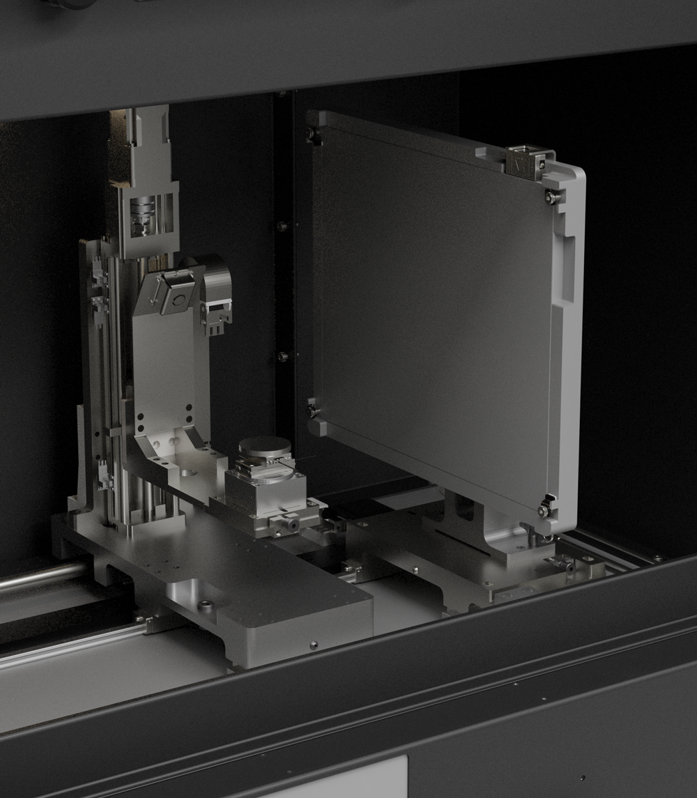
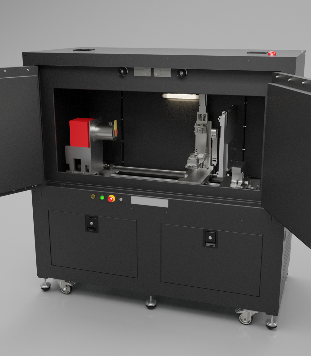
Re-configurable modular design
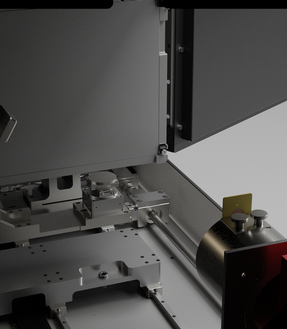
Sealed Type X-ray Source Up To 150 kV
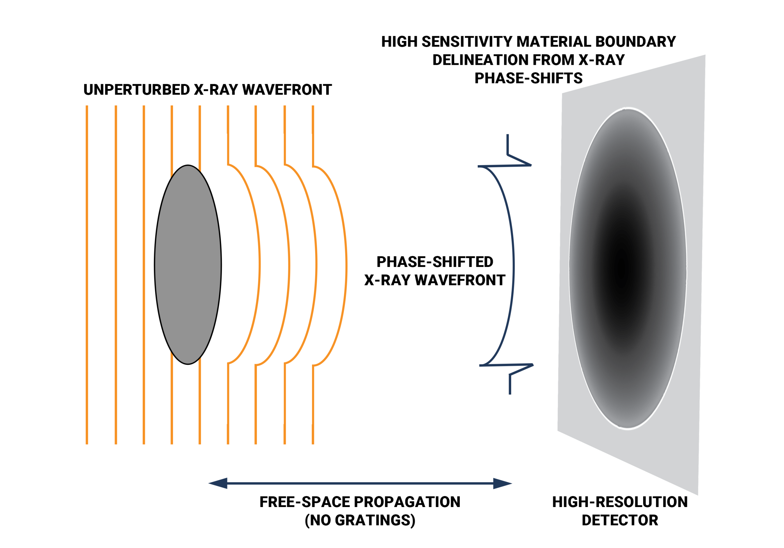
Propagation-based phase contrast Technology
X-ray Spot Size Down To 2 μm (110 kV max.)
Faster scan time
Literature
Product Gallery
inCiTe 2.0™: Phase Contrast and Spectral Imaging for Industrial and Research Excellence
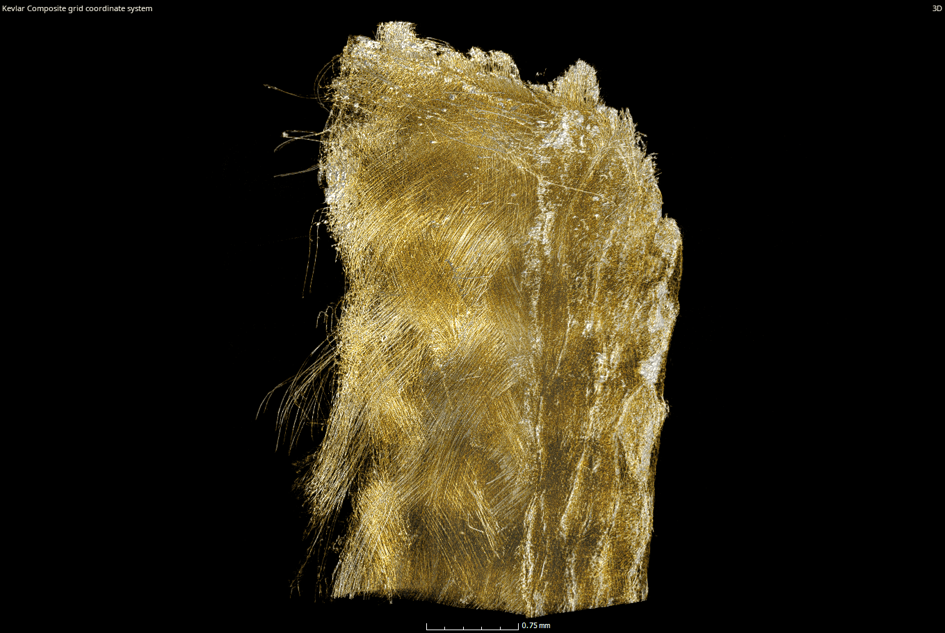
KA Imaging’s inCiTe 2.0 is a highly configurable 3D X-ray research workstation designed for complex material characterization and industrial NDT. By combining advanced phase contrast with spectral imaging, it enables precise multi-material differentiation in a single scan. Engineered for versatility, the inCiTe 2.0 features an expanded internal cabinet to accommodate custom in situ stages and a high-power 150kV source for denser samples. It offers a non-destructive 3D alternative to surface-level SEM, providing deep structural insights for biomechanics, aerospace, and advanced manufacturing.

Non-Destructive Testing (NDT)
Aerospace Component Inspection
Battery Analysis
Construction Materials Testing
Academic Research
Security Screening
Customer Stories
Phase Contrast Imaging as a Foundation
The inCiTe™ 2.0 system builds on the phase-contrast imaging capabilities established in the inCiTe™ 3D X-ray Microscope, which enhances visualization of low-density and weakly absorbing materials.
In inCiTe 2.0, phase contrast is combined with spectral imaging to extend analysis beyond structural detail, supporting improved differentiation of materials within complex and multi-material samples.
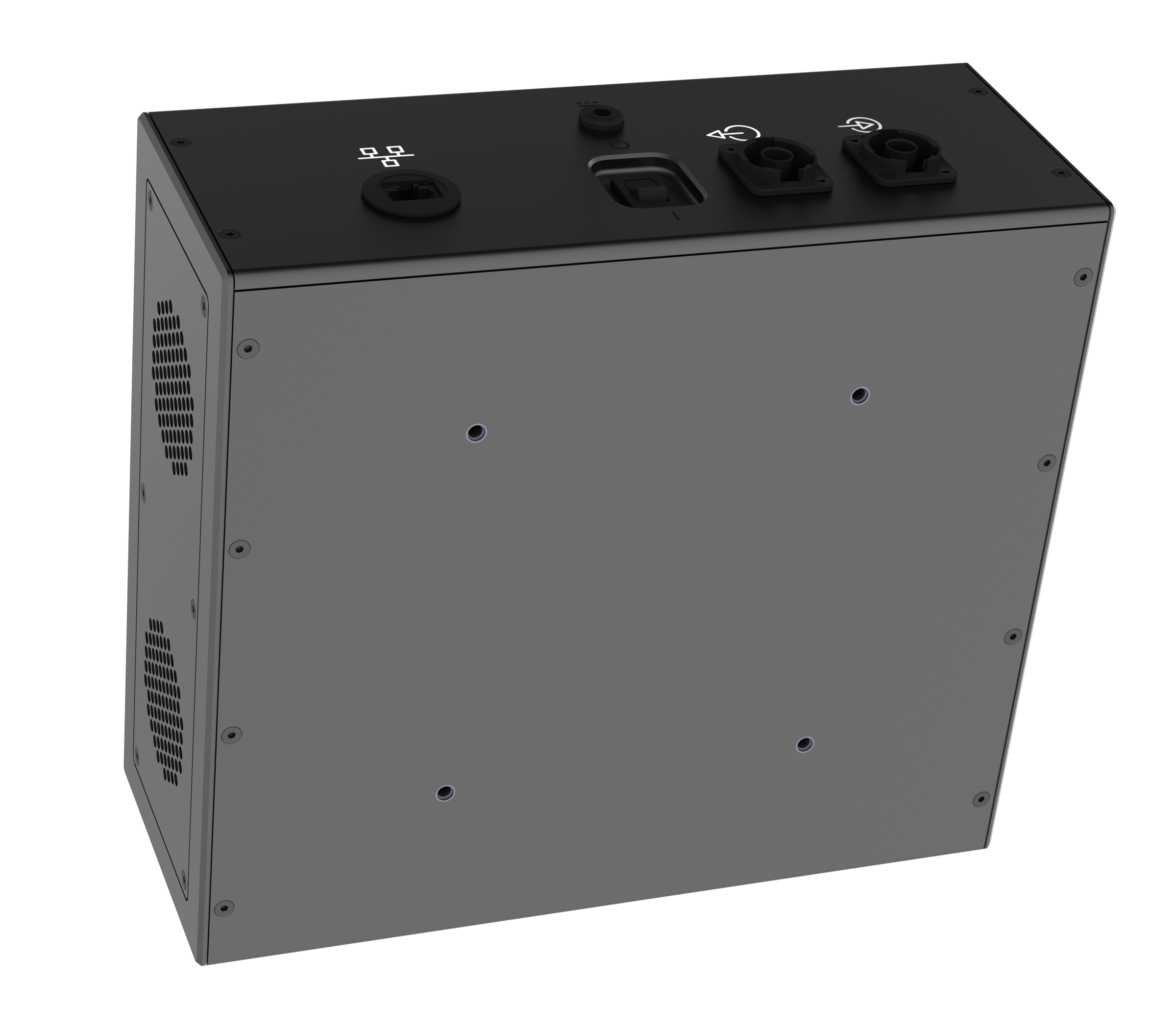
Detector Technology in a Multi-Modal System
inCiTe™ 2.0 incorporates the BrillianSe™ X-ray detector, which enables high spatial resolution and efficient X-ray detection through direct conversion.
For more details on the detector architecture and its role in phase-contrast imaging, see the inCiTe™ 3D X-ray Microscope.
Spectral Imaging for Material Differentiation
In addition to phase-contrast imaging, inCiTe™ 2.0 integrates spectral imaging capabilities through the Reveal™ platform, enabling further differentiation of materials within a single scan.
By capturing energy-dependent information at the detector level, spectral imaging supports the separation of materials with similar densities but different compositions, providing additional insight beyond structural detail alone.
For more information on spectral imaging and the Reveal™ platform, see the Reveal™ Platform.
Looking for dedicated soft-tissue clarity? See inCiTe
For applications focused on visualizing low-density and soft-tissue structures, the inCiTe™ 3D X-ray Microscope provides dedicated phase-contrast imaging in a compact, benchtop form factor. Designed to bring high-quality detail directly to your laboratory bench, it offers an efficient, space-saving solution for researchers who require high-resolution structural detail without the footprint of a large-scale floor station.
Explore inCiTe, our Phase Contrast 3D X-ray Microscope:
Which one is for me? inCiTe vs. inCITe 2.0
While both systems utilize our patented phase-contrast technology, they are designed for different operational environments. Paired with the BrillianSe detector, the inCiTe is our dedicated benchtop specialist, offering a compact, space-efficient footprint for researchers focused on high-resolution imaging of low-density materials (up to 110 kV).
In contrast, the inCiTe 2.0 is an expanded research workstation that can be configured with either the BrillianSe or Reveal detectors. It is built to accommodate high-power 150 kV sources and provides the expanded internal cabinet volume necessary for custom in situ stages and complex biomechanical rigs.
|
Feature / Capability
|
inCiTe (Phase Contrast)
|
inCiTe 2.0 (Spectral)
|
|---|---|---|
|
Dimensions |
Dedicated Phase Contrast clarity in a small footprint. |
Spectral imaging + high-load In Situ flexibility. |
|
Max X-ray Source |
110 kV |
Up to 150 kV |
|
Primary Technology |
Propagation-based Phase Contrast |
Phase Contrast + Spectral (Color) Imaging |
|
Material Specialty |
Low-Density Materials: |
Multi-Material Discovery: |
|
Key Advantage |
Maximum contrast for nearly "invisible" soft samples. |
Ability to "color-code" materials based on atomic number (Z). |
|
Imaging Modes |
Absorption and Phase Contrast. |
Absorption, Phase Contrast, and Spectral Material Decomposition. |
|
Sample Density |
Optimized for low to medium-density samples. |
High versatility; handles low-density to high-density industrial parts. |
|
Best For... |
Analytical Researchers: |
Biomechanical & Industrial Engineers: |
inCiTe Media Gallery
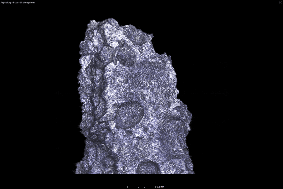
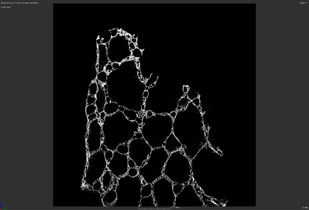
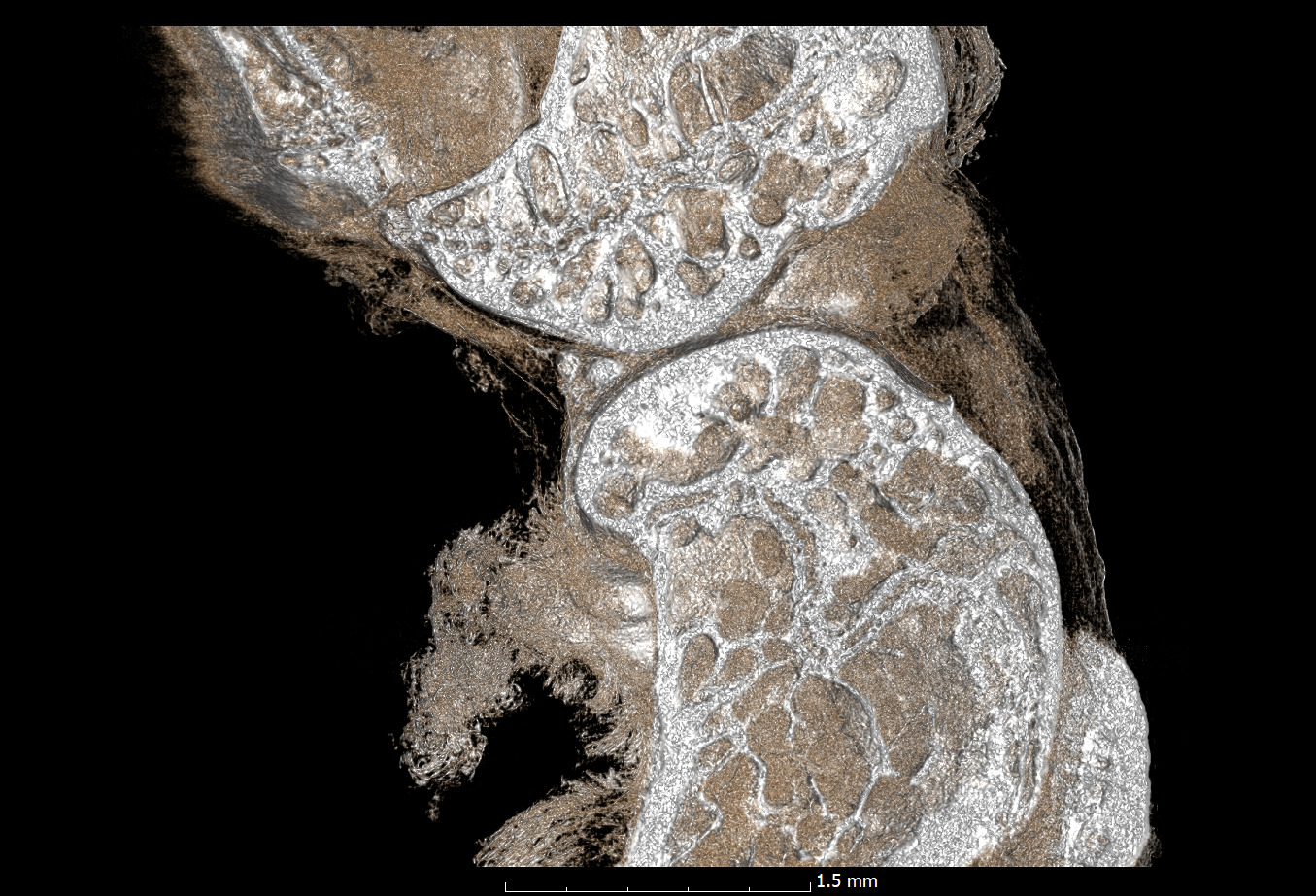
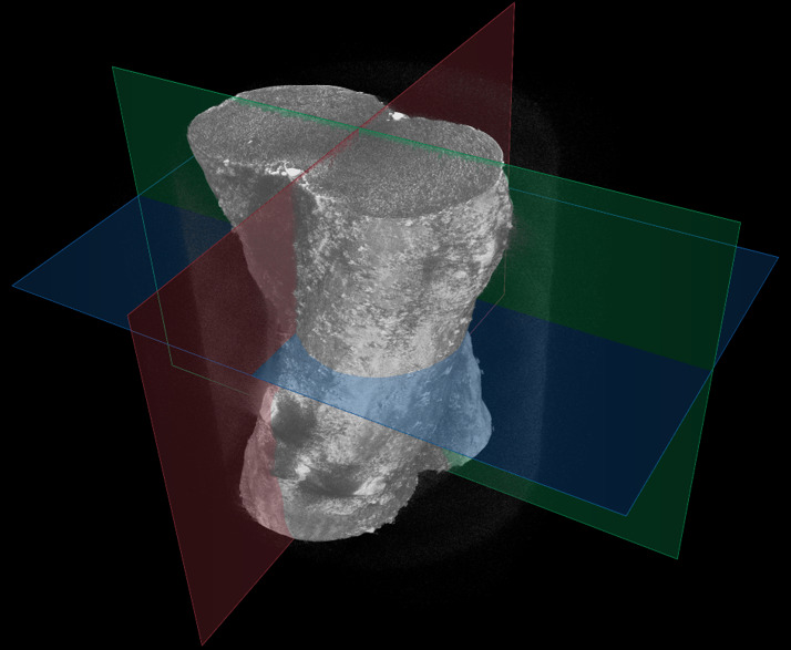
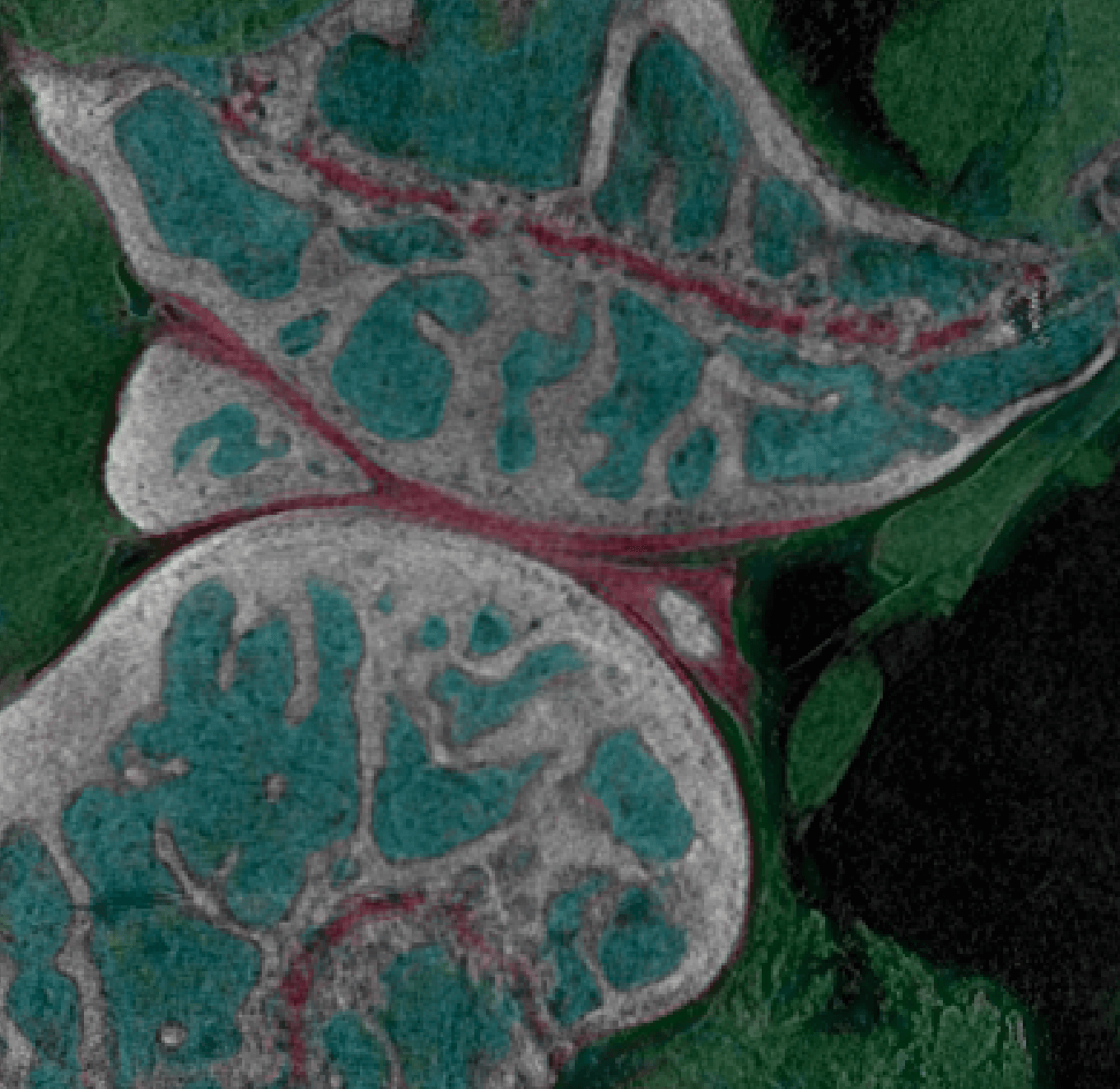
Images shown represent phase-contrast imaging capabilities. inCiTe™ 2.0 extends these capabilities with spectral imaging.
Latest blogs
Looking Beyond the Surface You’ve inspected the surface of a failed component and found nothing…
Biomedical research often requires scientists to visualize structures with vastly different X-ray attenuation properties within…
What Is Low Flux Imaging? Low flux imaging refers to imaging conditions where only a…
Original research
S. Abbaszadeh, CC Scott, O Bubon, A Reznik, KS Karim, “Enhanced detection efficiency of direct conversion X-ray detector using polyimide as hole-blocking layer,” Scientific Reports, December 2013
G.P. Lindberg, T. O’Loughlin, N. Gross, A. Mishchenko, A. Reznik, S. Abbaszadeh, K.S. Karim, G. Belev, B. Weinstein, “Photo-crystallization in a-Se layer structures: effects of film-substrate interface-rigidity,” Journal of Applied Physics, vol. 116, November 2014.
A. Parsafar, C. Scott, A. El-Falou, P. Levine, K.S. Karim, “Direct-Conversion CMOS X-Ray Imager with 5.6 μm × 6.25 μm Pixels,” IEEE Electron Device Letters, 36(5), pp. 481-3, May 2015.
C.C. Scott, A. Parsafar, A. El-Falou, P. M. Levine, K.S. Karim, “High Dose Efficiency, Ultra-high Resolution Amorphous Selenium/CMOS Hybrid Digital X-ray Imager,” IEEE International Electron Devices Meeting (IEDM) Technical Digest, December 2015.
Procházková, P., Kaiser, M., Stravová, Z., Petřík, M., Karim, K. S., Birch, Z., Comai, G. E., Corb, A., Tajbakhsh, S., Zikmund, T., & Kaiser, J. (2025, September). Revealing the architecture of eye muscles using cryogenic X-ray imaging [Oral presentation]. Tomography for Scientific Advancement (ToScA) Conference, France.
Related research
- Comparing conventional and phase contrast imaging
Gureyev, Timur Eugenievich, Sheridan C. Mayo, Damian E. Myers, Ya Nesterets, D. M. Paganin, A. Pogany, Andrew W. Stevenson, and S. W. Wilkins. “Refracting Röntgen’s rays: propagation-based x-ray phase contrast for biomedical imaging.” Journal of Applied Physics 105, no. 10 (2009): 102005.
Krenkel, Martin, Mareike Töpperwien, Christian Dullin, Frauke Alves, and Tim Salditt. “Propagation-based phase-contrast tomography for high-resolution lung imaging with laboratory sources.” AIP Advances 6, no. 3 (2016): 035007.
Olivo, A., and E. Castelli. “X-ray phase contrast imaging: From synchrotrons to conventional sources.” La Rivista Del Nuovo Cimento 37 (2014): 467-508.
Bravin, Alberto, Paola Coan, and Pekka Suortti. “X-ray phase-contrast imaging: from pre-clinical applications towards clinics.” Physics in Medicine & Biology 58, no. 1 (2012): R1.
Bidola, P., K. Morgan, M. Willner, A. Fehringer, S. Allner, F. Prade, F. Pfeiffer, and K. Achterhold. “Application of sensitive, high‐resolution imaging at a commercial lab‐based X‐ray micro‐CT system using propagation‐based phase retrieval.” Journal of Microscopy 266, no. 2 (2017): 211-220.
- Propagation
Wilkins, S. W., T. Ei Gureyev, D. Gao, A. Pogany, and A. W. Stevenson. “Phase-contrast imaging using polychromatic hard X-rays.” Nature 384, no. 6607 (1996): 335-338.
Mayo, Sheridan C., Andrew W. Stevenson, and Stephen W. Wilkins. “In-line phase-contrast X-ray imaging and tomography for materials science.” Materials 5, no. 5 (2012): 937-965.
- Gratings
Pfeiffer, Franz, Timm Weitkamp, Oliver Bunk, and Christian David. “Phase retrieval and differential phase-contrast imaging with low-brilliance X-ray sources.” Nature physics 2, no. 4 (2006): 258-261.