
Critical Care & Bedside Imaging
Improving Diagnostic Insight at the Patient’s Bedside

Portable chest X-ray imaging plays an essential role in intensive care environments where patients often cannot be transported safely to the radiology department. Bedside imaging allows clinicians to monitor disease progression, verify placement of medical devices, and assess complications directly in the ICU.
KA Imaging provides advanced X-ray inspection technologies designed for laboratory and at-line quality control environments, enabling detailed internal inspection of components, assemblies, and materials across a wide range of industries.
Spectral X-ray imaging expands the information available during bedside imaging. KA Imaging’s Reveal™ platform uses SpectralDR® technology to generate conventional, bone, and soft-tissue images from a single exposure while maintaining standard portable radiography workflow.
Solutions for Critical Care Imaging
KA Imaging’s Reveal™ platform enables spectral imaging during routine portable chest X-ray exams performed in intensive care units and other bedside environments.
Relevant solutions include:
Clinical Evidence in ICU Environments
Clinical studies evaluating spectral imaging in intensive care environments have shown promising results.
Clinical studies using the Reveal 35C have suggested:
67%
of radiologists reported improved image quality⁹
67%
reported faster or equivalent reading times⁹
37%
Up to 37% decrease in the number of chest CT scans for ICU patients⁹
Increased
Increased diagnostic among intensivists⁹
These results suggest that spectral imaging can provide additional diagnostic insight while maintaining the efficiency of routine portable radiography.
Why Spectral X-ray Imaging Matters in Critical Care
Conventional radiography compresses all anatomical information into a single image. In complex clinical environments, overlapping structures may obscure clinically relevant findings.
Spectral X-ray imaging separates bone and soft-tissue information from a single exposure, producing multiple image outputs while maintaining the same workflow as standard radiography.
For clinicians, this means:
- additional imaging information without additional exposures
- no change to portable radiography workflow
- improved visualization in complex bedside environments

Why choose dual-energy (Spectral) X-ray?
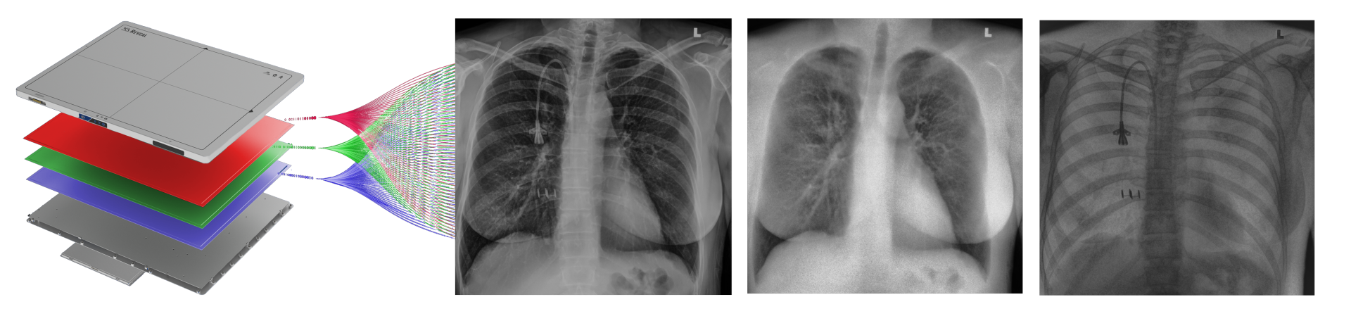
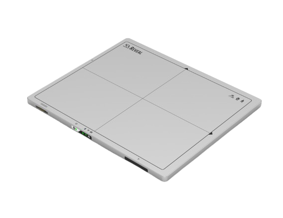
Reveal™ 35C detectors use SpectralDR® technology to acquire multiple spectral images from a single X-ray exposure.
Each exposure produces:
- a conventional digital radiography image
- a bone image
- a soft-tissue image
These images allow clinicians to separate overlapping anatomical structures and visualize areas that may be less visible in conventional radiographs.
Unlike traditional dual-energy imaging techniques that require multiple exposures, SpectralDR captures these images simultaneously with a single exposure, eliminating motion artifacts and maintaining the same clinical workflow as standard portable radiography.
Key Clinical Scenarios in Critical Care
Lines and Tubes Verification
Spectral imaging can improve visualization of catheter tips and other medical devices by separating bone structures from surrounding soft tissue.


Pneumothorax Evaluation
Soft-tissue images can reduce rib overlap and may improve visualization of pneumothorax during bedside chest X-ray examinations.
Pneumonia and Lung Lesions Monitoring
Spectral imaging can enhance visualization of lung opacities by separating bone from soft tissue, potentially improving evaluation of pneumonia and other lung conditions.


Trusted by Users
Patrik RogallaRadiologist
In the first 34 patients, Reveal™’s dual energy capabilities have already helped find pneumonia with higher confidence. We are continuing to image and will be adding a second system to increase our imaging capacity.
Sandeep RongheMedical Imaging Technologist
I think the patients with the least mobility will be impacted the most. If we can take the imaging test to them, that makes a really big difference. Overall, just the sheer dose level being cut down to significant amount of dose makes it the kind of imaging technology that is worth to have.
Carla GirolamettoDirector of Research, Innovation and Clinical Trials, Grand River Hospital, Kitchener
A preliminary analysis comparing the six weeks preceding the pilot period and the 6 weeks in which KA Imaging’s 35C Reveal™ detector was in use demonstrates a decrease in both the total number of portable chest x-rays as well as chest CTs for patients admitted to ICU.
Related Applications

Enhance Bedside Imaging in Critical Care
Learn how spectral X-ray imaging can improve diagnostic insight during portable radiography in intensive care environments.
Latest blogs
Global air cargo volumes continue to rise year after year, placing growing pressure on airport…
For critically ill or post-surgical patients, transporting them to a dedicated radiology department is a…
Medical imaging plays a critical role in diagnosis, treatment planning, and patient monitoring. The goal…