
Musculoskeletal radiography is widely used to evaluate bones, joints, and surrounding structures. X-ray imaging plays a central role in assessing fractures, degenerative changes, and other skeletal conditions.
However, interpretation of conventional radiographs can be challenging when overlapping anatomical structures obscure clinically relevant features. Soft tissue, overlapping bones, and complex joint anatomy may reduce visibility of certain structures.
Spectral X-ray imaging expands the information available in routine musculoskeletal radiography. KA Imaging’s Reveal™ platform uses SpectralDR® technology to generate multiple image types from a single exposure while maintaining the same workflow and radiation exposure as conventional radiography.
These additional images allow clinicians to view bone and soft-tissue structures separately, which may improve visualization of skeletal anatomy.
Solutions for Musculoskeletal Imaging
Reveal™ spectral imaging solutions can be used during routine musculoskeletal radiography exams.

Relevant solutions include:
Key Clinical Scenarios
Skeletal Structure Visualization
Bone images generated by spectral imaging emphasize calcified structures, allowing clinicians to review skeletal anatomy with reduced soft-tissue overlap.
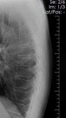
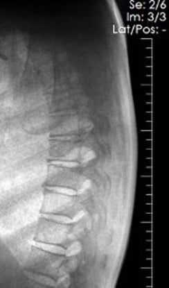
The Bone image is more adept at presenting the bony skeleton in a manner that delineates it better than a conventional DR image. By displaying only on the areas of highest subject density, the SpectralDR™ technology yields improved visualization of both healthy and diseased bone.
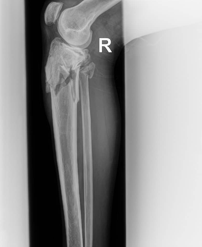
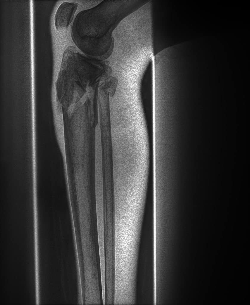
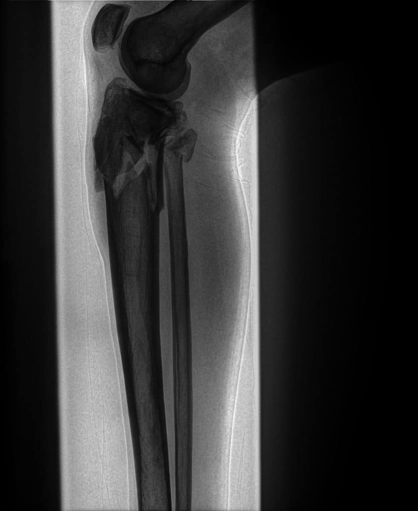
SpectralDR bone images emphasize skeletal structures, allowing clearer visualization of the fracture compared with the conventional radiograph.
Evaluation of Calcified Structures
Spectral imaging highlights calcified structures within the musculoskeletal system, allowing clinicians to examine bone anatomy using multiple image types generated from a single exposure.
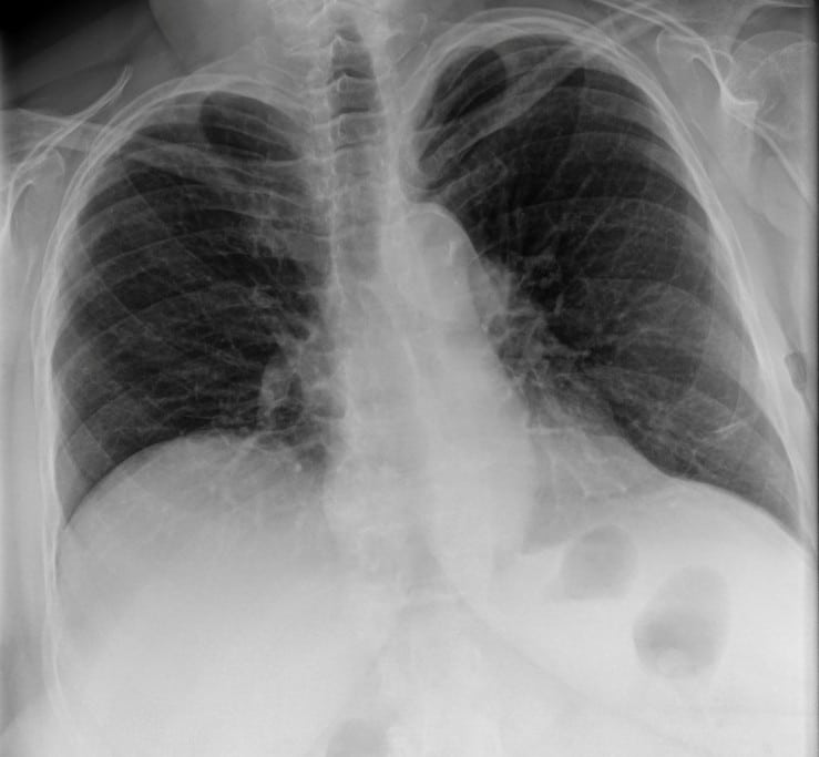
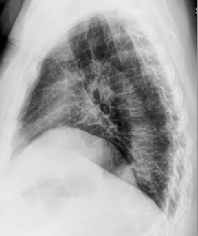
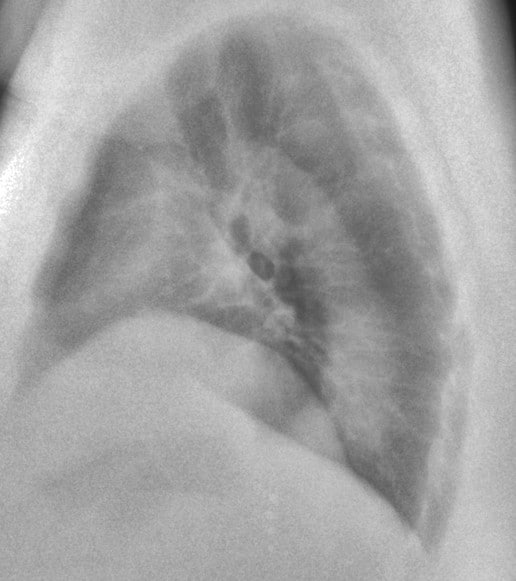
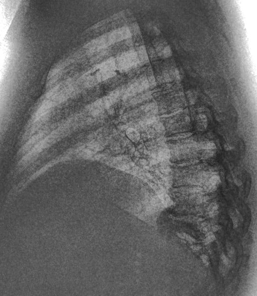
This 67-year old female patient presented to the ER with shortness of breath, in conjunction with chronic pain in her posterior thoracic region upon taking a large breath in. The reading radiologist noticed a suspicious mass-like opacity in the Lateral view. The radiologist stated that, thanks to the DE images, they could they tell the opacity wasn’t a lung mass, but instead far less concerning bony outcroppings from the thoracic vertebrae.
Trusted by Users
Dr. Methuselah KorirOrthopedic Surgeon
The SpectralDR provides a depth of detail far beyond that of standard X-rays, allowing for the diagnosis of the subtlest pathologies that would otherwise be missed. Being able to clearly distinguish fracture fragments and soft tissue involvement has fundamentally improved my surgical planning and diagnostic confidence.
Spectral Imaging for MSK
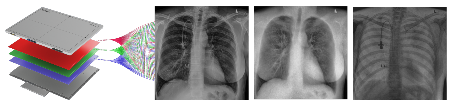
Reveal™ detectors generate three images from a single X-ray exposure:
• conventional radiograph
• bone image
• soft-tissue image
Bone images emphasize calcified structures and skeletal anatomy, while soft-tissue images reduce the visual impact of bone structures.
This separation allows clinicians to examine musculoskeletal anatomy from different perspectives using a single radiography exam.
Why Spectral X-ray Matters
In conventional radiography, bone and soft-tissue information are captured in a single image. In complex musculoskeletal anatomy, this overlap can make interpretation more challenging.
Spectral imaging separates bone and soft-tissue information from a single exposure, producing multiple image outputs that provide additional perspectives on anatomical structures.
This separation can help clinicians:
• visualize skeletal structures with reduced soft-tissue overlap
• evaluate bone anatomy in complex joints
• review musculoskeletal radiographs using multiple image types
All while maintaining the same workflow as standard radiography.
Related Applications
Emergency & Trauma Imaging
Chest Imaging
Critical Care & Bedside Imaging

Expand the Value of Musculoskeletal Radiography
Learn how spectral X-ray imaging can provide additional imaging insight during routine musculoskeletal exams.
Latest blogs
Imaging plays an important role in monitoring patient health and supporting clinical decision-making in intensive…
Tuberculosis remains one of the world’s deadliest infectious diseases. Currently, the critical challenge has shifted…
In 2015, KA Imaging was founded with one major goal: to redefine what X-ray technology…