Entries by Fernanda Fraga

How SpectralDR X-Ray and Micro-CT Technology Revolutionizes the Imaging of Aerospace Composite Materials

In the aerospace industry, ensuring the integrity and safety of components is absolutely crucial. With the high costs and vital importance of maintaining structural integrity, the industry has turned to advanced imaging technologies to meet these needs. X-ray and Micro-CT technology have emerged as revolutionary tools that provide extraordinary insights into the internal structures and potential flaws of aerospace parts. These technologies enable more accurate and comprehensive assessments, essential for maintaining the highest standards of safety and performance in aircraft and spacecraft. This blog explores how the innovative solutions from KA Imaging, such as the Reveal 35C and inCiTe 3D X-ray microscope, are transforming aerospace non-destructive testing (NDT) by delivering detailed imaging that goes beyond what traditional methods can offer.

Importance of Quality Testing and NDT in Aerospace Industry

As an industry with safety at the forefront of their operations, non-destructive testing methods are pivotal to the aerospace industry. Aerospace components can experience many non-visible defects, and depend on NDT to ensure the safety of aircrafts, spaceships, and more. Destructive testing methods are not often utilized for aerospace, as aerospace components are extremely costly to produce. NDT gives aerospace companies the opportunity to perform quality testing while still maintaining the functionality of aerospace components, such as an aircraft’s elevator. This ensures that, if the component is considered viable, the components can continue to be used in everyday operations. Throughout the years, aerospace has prominently utilized several NDT methods to diagnose manufacturing defects. 

Testing Methods in Aerospace NDT

There are many NDT methods that are commonly used within the aerospace industry: 

  • Ultrasonic Testing (UT): Ultrasonic Testing can be effectively performed on metal materials such as steel and iron. It sends short ultrasonic waves through a specimen to detect any audible discrepancies. Although UT is most functional with metal materials, it is also viable to be used with concrete or wood materials, although with weaker sound detection. 
  • Magnetic Particle Inspection (MPI): Magnetic Particle Inspection is used with ferromagnetic materials, most commonly iron, copper, and nickel. It can also be used on certain alloys or compounds that contain trace amounts of ferromagnetic materials. It determines defects like cracks beneath the surface of a specimen by running a magnetic current through it. The magnetic particles will accumulate at the site of cracks or discrepancies. 
  • Fluorescent Penetrant Inspection (FPI): Fluorescent Penetrant Inspection is a NDT technique designed to find surface defects in materials like metals or plastics, or any material that is non-porous. It involves applying a low viscous fluorescent dye to the surface of a specimen. The dye will seep into any surface cracks or discrepancies.  
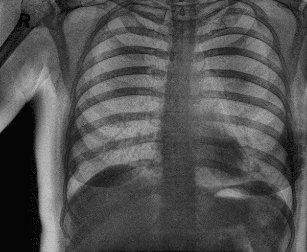
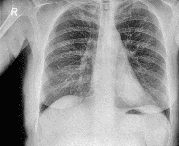
  • Radiographic Testing (RT): Radiographic Testing is what KA Imaging specializes in. RT uses X-rays or gamma rays to create an image of the inside of a specimen. These images are then examined by specialists to identify abnormalities in structure and/or general defects. It is similar in practice to medical radiography, but instead examining objects and materials. 

What is Micro-CT Radiographic Non-Destructive Testing? 

Micro-CT, or micro-computed tomography, is an advanced imaging technique that produces high-resolution, three-dimensional images. It creates images of an object’s internal structure without causing significant damage or requiring dissection. It is essentially a scaled-down version of the standard computed tomography (CT) scans used in medical imaging, providing much finer detail. Micro-CT is employed extensively in scientific research, material science, biology, geology, and industrial non-destructive testing. In micro-CT, the object itself is rotated 360 degrees. As it rotates, X-rays pass through it from various angles, producing a three-dimensional image of the object. 

What is SpectralDR technology for Non-Destructive Testing?

KA Imaging developed a patented cutting edge Dual Energy Subtraction technology known as SpectralDR. SpectralDR is unique from other DES technology because it acquires multiple energies in one X-ray exposure while not requiring increased radiation doses. The standard radiation dose needed for the average DR is all that is needed for Reveal’s technology to produce crisp imaging. Additionally, this imaging can be executed with no distortion or errors caused by motion. 

Triple Stacked Layer Design

Single exposure dual-energy subtraction X-ray – our SpectralDR technology – is only possible because of Reveal’s distinctive triple stack design. Thanks to SpectralDR, our mobile X-ray detector is capable of simultaneously acquiring three images with only one exposure. The technology is capable of producing separated imaging on bone or soft tissue, improving clarity and reducing the chances of important visualization being obstructed. 

This advanced technology enhances the identification of different materials and improves the detection of cracks, corrosion, and other critical defects in aerospace components. With its ability to produce clearer and more detailed images, SpectralDR ensures the structural integrity and reliability of aerospace parts, contributing to safer and more efficient aircraft maintenance and manufacturing processes.

What sets KA Imaging’s NDT products apart

Since 2015, KA Imaging has remained a trailblazer in the world of X-ray imaging. We streamline radiographic technology, creating various unique products in the realm of micro-computed tomography, high-efficiency X-ray area detection, and multi-energy spectral separation detection.

KA Imaging supports aerospace use cases with our Reveal 35C X-ray detector and inCiTe 3D X-ray microscope. Reveal 35C is the first portable dual-energy single exposure X-ray detector, capable of single exposure dual-energy imaging. Reveal 35C generates three image types, traditional dr, and then two other images which highlight the low-density or high-density materials. This allows for advanced material separation for more comprehensive inspection. InCiTe pairs with Reveal 35C to offer phase contrast and micro-CT. InCiTe can provide enhanced visualization of low-density materials with its superior contrast, as well as producing three-dimensional images with its micro-CT technology. InCiTe uses propagation based X-ray imaging for enhanced visualization of objects with poor x-ray absorption.

KA Imaging’s products offer a distinct and customizable visual representation of an object’s structure. Radiographic inspectors can use Reveal 35C’s software to customize between focused imaging prioritizing high or low density material detection, or traditional imaging. Additionally, Reveal 35C uses single exposure, removing the appearance of motion artifacts which can disrupt visualization.  

aerospace ndt conventional dr
aerospace ndt low z application
aerospace ndt high z application

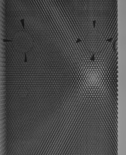
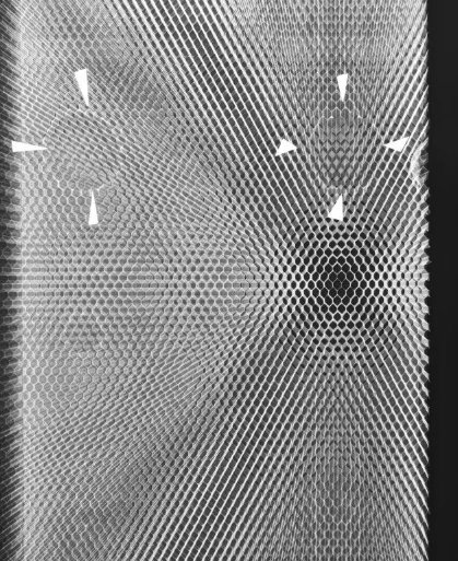
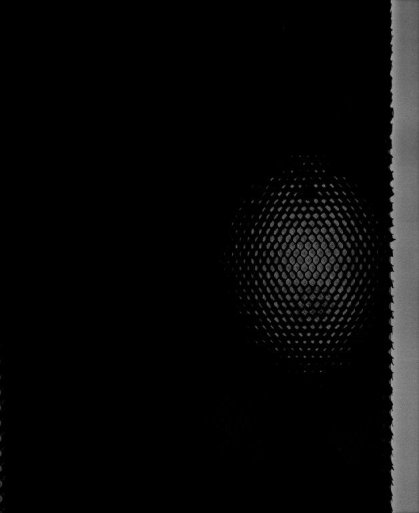
In the above image of a titanium composite wing, you can see a conventional DR image on the left paired with a low Z application in the middle, and high Z application on the right. Changes in matter caused by increasing or decreasing Z can affect photoelectric absorption 

With technology like Reveal and inCiTe, aerospace quality testing can reliably produce more comprehensive imaging of structurally complex components. While other NDT methods give a partial impression of object discrepancies, KA’s radiographic technology offers clear, three-dimensional imaging that leaves a permanent record in the form of radiographic film or digital images. This record allows images to be repeatedly inspected for reinforced accuracy when detecting discrepancies. 

Learn more about Reveal Technology. 

Learn more about inCiTe 3D Technology. 

Revolutionizing Aerospace NDT with KA Imaging 

The integration of X-ray and Micro-CT technology into aerospace non-destructive testing represents a significant advancement in the industry. By offering high-resolution, three-dimensional imaging, these techniques provide a more complete and nuanced understanding of material integrity compared to traditional methods. KA Imaging has been at the forefront of this revolution, with innovative products like the Reveal 35C and inCiTe 3D X-ray microscope setting new benchmarks for accuracy and efficiency in aerospace NDT. These technologies not only enhance the detection of potential defects but also ensure that components can be safely and effectively used, thereby reducing costs and improving operational reliability. As the aerospace industry continues to evolve, the adoption of advanced imaging techniques will play an increasingly crucial role in maintaining the highest levels of safety and performance. Explore KA Imaging’s groundbreaking solutions today to find out how Reveal and inCiTe can elevate your aerospace quality testing processes.

How Reveal 35C Empowers CXR For Pneumonia Diagnosis

Since X-rays first began examining soft tissue in the early 20th century, CXR has played a pivotal role in diagnosing pneumonia. Early radiographs were fairly rudimentary compared to modern standards, but they allowed physicians to see pneumonia-associated changes in lung fields. In the last 100 years since its discovery, X-ray lacked any major technological advancements until the introduction of SpectralDR, developed by KA Imaging. Our team at KA Imaging has remained trailblazers in the radiography landscape, innovating imaging technology to new heights. This article explores the importance of enhanced CXR for pneumonia, and how KA Imaging’s patented Reveal 35C X-ray Detector with SpectralDR technology innovates the standards for accurate CXR. 

The Importance Of Accurate CXR For Pneumonia

Pneumonia often presents with infiltrates, which are areas of increased density (opacity) in the lung tissue due to infection and inflammation. What makes enhanced CXR for pneumonia so important is that it helps not just diagnose, but also determine the extent and location of the pneumonia. This includes identifying whether it is affecting a single lobe (lobar pneumonia), multiple lobes (multilobar pneumonia), or diffusely spread across the lungs (bronchopneumonia). This increased level of knowledge in the pneumonia’s severity has a major impact in how medical personnel administer life-saving treatment. Moreover, radiographic visualization is sometimes able to detect complications caused by pneumonia, such as empyema or abscess formation. 

Based on the CXR findings, medical personnel can decide whether further testing is necessary. Our research has found that there are many cases in which our X-ray technology alone can identify pneumonia in patients. Diagnosis solely using a single X-ray examination when possible is beneficial for both patients and medical facilities. 

Symptoms of pneumonia can overlap with other chest conditions such as congestive heart failure, pulmonary edema, lung abscesses, or malignancies. This is where intensive CXR visualization and accuracy becomes most important. Accurate CXR helps differentiate pneumonia from these other possibilities through visualization of unique, characteristic lung patterns. 

Reveal with SpectralDR technology: The World’s First Portable Dual-Energy Detector

As CXR plays such an important role in pneumonia diagnosis, radiographic technology developers have worked on improving aspects such as image quality or technology deployment (like with mobile systems). But essentially, digital radiography has remained fairly the same. With KA now introducing revolutionary imaging technology that goes beyond just image quality and resolution, various conditions that are visualized in radiography are easier to visualize, examine, and therefore, diagnose. Quick diagnosis unlocks the potential for even faster medical treatment. 

KA Imaging’s mission is to empower limitless innovation for radiographic imaging, simplifying and supporting medical testing efforts to improve detailed identification of life-threatening conditions, including pneumonia. 

The Reveal 35C X-ray Detector

KA Imaging developed the world’s first portable single exposure dual-energy detector, the Reveal 35C X-ray Detector. This patented technology is called SpectralDR. Reveal 35C is capable of improving the visualization of many conditions through radiographic imaging, but is most known for its good results achieved in detecting signs of pneumonia with CXR. Let’s dive into how Reveal 35C’s technology enhances radiographic visualization. 

Dual-Energy Subtraction (DES): The Old Ways vs. SpectralDR technology

Old approaches of DES technology involve taking two separate X-ray exposures at different energy levels (usually a high-energy and a low-energy exposure). The difference in absorption of X-rays by various tissues at these two energy levels provides a basis for separating images of different tissue types.

KA Imaging developed a patented cutting edge DES technology known as SpectralDR. SpectralDR is unique from other DES technology because it acquires multiple energies in one CXR scan while not requiring increased radiation doses. The standard radiation dose needed for the average CXR is all that is needed for Reveal’s technology to produce crisp imaging. Additionally, this imaging can be executed with no distortion or errors caused by motion. 

Triple Stacked Layer Design

Single exposure dual-energy subtraction X-ray – our SpectralDR technology – is only possible because of Reveal’s distinctive triple stack design. Thanks to SpectralDR, our mobile X-ray detector is capable of simultaneously acquiring three images with only one exposure. The technology is capable of producing separated imaging on bone or soft tissue, improving clarity and reducing the chances of important visualization being obstructed. 

How Reveal Technology Highlights Visible Signs Of Pneumonia

Thanks to its innovative 3-in-1 design, Reveal 35C is capable of capturing diverse imaging of the subject with just a single exposure. It can capture images of bones and tissues, together and individually. This means that doctors will obtain imaging that focuses both on the soft tissue, and alternatively the bone surrounding it, in the case of a CXR for pneumonia, the ribs and lungs. With Reveal 35C, doctors can effectively capture and visually differentiate lung nodules, ribs fractures, and even foreign objects within the body with ease. 

cxr for pneumonia soft tissue spectraldr imaging
cxr for pneumonia rib bone spectral dr imaging
cxr for pneumonia combined imaging of bone and soft tissue

On the left, the soft tissue is visible with no rib imaging to obstruct it. In the middle image, the Reveal 35C produces distinct exposure of the rib bones. 

Reveal 35C’s 3-in-1 technology provides three distinct photos of the subject, a standard image of the ribs and lungs, a photo highlighting just the ribs, and a photo visualizing beyond the ribs so that only the lungs are visible.

In a paper presented at RSNA in 2021, the Reveal 35C technology was shown to detect 33% more pneumonia cases (including COVID-19) than traditional X-Ray. The paper presents the initial results of an ongoing clinical trial that is being carried out in Toronto (Canada). 

With Reveal 35C, medical personnel can overcome visual obstructions caused by bone, in this case the ribs, to get a better view of the soft tissue, the lungs. This makes it possible to examine signs of pneumonia that would otherwise be obstructed and unviewable from behind the ribs. This has allowed for quicker identification and improved accuracy in diagnosis when performing CXR for pneumonia. As previously mentioned, this not only applies to diagnosis, but also examination of the pneumonia’s severity and appearance of side effects. The quicker the pneumonia and its side effects are identified and diagnosed, the quicker medical personnel are able to administer adequate treatment. If a single x-ray can identify pneumonia and its side effects, medical personnel can potentially avoid the need for further imaging, speeding up treatments and saving on cost, time, and resources. 

Reveal 35C is the first single exposure dual-energy X-ray detector to deliver a portable imaging solution, only sold by KA Imaging. Mobile imaging technology is becoming a necessity for medical personnel, as it allows more flexibility in emergency situations, long-term care homes, or when examining patients who cannot move easily like in the ICU. Medical facilities aim to limit the transportation of highly infectious patients in order to avoid contaminating additional spaces within the facility. Mobile X-ray imaging allows for doctors to assess patients from their bedside, reducing the risk of constant movement of patients with contagious conditions, such as COVID-19. Reveal 35C is the first X-ray detector capable of capturing single exposure dynamic imaging in mobile use cases, expanding the capabilities of doctors and the comfort they can provide for their patients. 

At KA Imaging, our team is focused on innovating imaging technology with innovation of our phase contrast and Spectral DR features. Learn more about Reveal 35C’s capabilities, how to purchase a Reveal 35C for your medical facility, and what’s next for KA Imaging’s X-ray technology.

The importance of accurate CXR in identifying the location, extent, and complications of pneumonia cannot be overstated. KA Imaging’s Reveal 35C, with its patented Dual-Energy Subtraction (DES) and triple-stacked layer design, marks a revolutionary advancement in CXR for pneumonia. By providing clear, obstruction-free images with a single exposure, Reveal 35C aids precise and speedy diagnosis, critical for effective patient care. Its portability further enhances its utility, especially in point-of-care settings where rapid, accurate diagnostics are paramount. Investing in innovative solutions like the Reveal 35C not only improves diagnostic accuracy for pneumonia but also elevates the overall standards of medical care, ultimately saving lives and enhancing patient treatment.

,

Bringing ‘inCiTe’ To NDT: How KA Imaging Perfects NDT

In the realm of industrial safety and quality assurance, Non-destructive Testing (NDT) emerges as a cornerstone methodology, crucial for inspecting and ensuring the integrity of materials without compromising their usability. Known under various nomenclatures such as Non-destructive Examination (NDE) and Non-destructive Inspection (NDI), this technique plays a pivotal role across a diverse range of sectors—from aerospace to automotive. It essentially allows experts to detect potential problems before they escalate into costly or hazardous failures. This article delves into the significance of scientific imaging for NDT and introduces a groundbreaking advancement in this field with KA Imaging’s inCiTe and BrillianSe technology. 

X-Ray NDT and Quality Control

Non-destructive testing (NDT) plays a vital role in quality control, as it can be used to evaluate flaws or discrepancies in objects without compromising the functionality. This means that it can effectively study the health of various components over time and detect issues before an actual malfunction occurs. Typically, this can be achieved without any significant damage to the sample. 

Here are a few of the industries that benefit most from non-destructive testing (NDT): 

  • Security: This is where NDT is most commonly known, it can be used to screen luggage or other items
  • Automotive: NDT is used to inspect the health of various automotive parts, including EV batteries.
  • Additive manufacturing: NDT is used to inspect the quality of 3D printed materials
  • Aerospace: NDT is used to inspect aircraft components for safety.
  • Construction: NDT can evaluate the integrity of buildings or pipes, detecting cracks and defects.

Although these are not all the industries that actively utilize NDT, they are among the most prominent. Overall, NDT has a near endless variety of uses in an equally endless number of industries. 

Introducing KA Imaging’s inCiTe Technology

Since 2015, KA Imaging has developed innovative X-ray imaging technology and systems for use in multiple industries. 

KA Imaging offers inCiTe, the first commercial X-ray CT system to utilize BrillianSe technology to utilize advanced technology for detailed three-dimensional imaging. inCiTe employs Micro-CT to analyze each layer of an object meticulously, enhancing the ability to spot smaller discrepancies as soon as they become visually apparent.

The exceptional performance of inCiTe is primarily due to its propagation-based phase contrast, which naturally increases the contrast within samples based on X-ray phase changes. When paired with the innovative BrillianSe X-Ray Detector, inCiTe achieves remarkable results in quality assurance, making it capable of highlighting quality issues in a wide array of material samples.

BrillianSe Novel Detector Technology

BrillianSe is the latest innovation in X-ray detection technology, functional for high-brilliance imaging. It is a direct conversion detector, forgoing the need to first convert X-ray photons to be visible. The detector utilizes high spatial resolution amorphous selenium (a-Se) and propagation-based (grating less) in-line phase-contrast. KA Imaging developed the detector with a unique hybrid of a-Se in addition to complementary metal oxide semiconductor (CMOS) sensors. BrillianSe is the world’s highest spatial resolution X-ray detector, and is exclusively developed and patented by KA Imaging.

inCiTe And BrillianSe For NDT

  • Low-density Material Phase Contrast: The BrillianSe X-ray Detector can be applied for low-density material phase contrast, providing higher resolution images than X-ray technology without similar detection equipment. In the biomedical industry, BrillianSe can be employed to analyze the composition of low-density pharmaceuticals, such as tablets.
  • Synchrotron Micro-nano CT: BrillianSe pairs well with both micro-computed tomography (micro-CT) and nanotomography (nano-CT) technologies. 

How inCiTe and BrillianSe Improves Non-Destructive Testing (NDT)

BrillianSe serves as an innovation for non-destructive testing with a hybrid of a-Se and CMOS technology. The photoconductor a-Se enhances sensitivity when imaging low-density materials, making it capable of examining fragile samples without the risk of significantly damaging them. It is the only direction conversion material to be used in commercial flat-panel X-ray imaging (FPXI). Combined with CMOS, which allows for direct detection without loss of resolution, BrillianSe utilizes the advantages of both CMOS and a-Se technology. CMOS is also famous in the imaging industry for its ability to inspect with lower power consumption and noise levels than other detection sources. 

Since some materials have poor x-ray adsorption compared to others, inCiTe’s phase contrast serves as a tool to achieve heightened visibility of a sample with low absorption. Along with the BrillianSe phase contrast edge enhancement, this technology creates a distinct contrast on images, making the intricacies of a specimen’s composition more visible. This expands the library of materials that scientific imaging can effectively inspect, no longer being limited by a material’s level of absorption. With the inCiTe microscope and BrillianSe detector, discrepancies are easier to locate and decipher through its heightened contrast. The state-of-the-art technology highlights areas that may not appear as distinctly, or at all, with normal x-ray usage. 

Let’s take a look at the inCiTe microscope and BrillianSe detector in action:

BrillianSe image (left) of 18650 lithium ion battery, and BrillianSe image with an included 0.5mm aluminum filter (right) showing more bent cathodes and anodes. 

Using inCiTe’s phase contrast with BrillianSe’s phase contrast edge enhancement on this lithium ion battery vastly enhances the clarity of the sample. Other commercial detectors on the market may not be able to process materials of this size as effectively without the resolution and phase contrast inCiTe and BrillianSe offers.

KA Imaging’s inCiTe and BrillianSe technology marks a revolutionary stride forward in the domain of scientific imaging for non-destructive testing. They offer unmatched clarity and enhanced detection capabilities that boost safety and efficiency across numerous industries. Where high performance meets affordability, inCiTe and BrillianSe sticks the landing. With its unique blend of amorphous selenium (a-Se) and CMOS sensor technology, BrillianSe facilitates better visualization and identification of discrepancies, even in low-density materials. If you’ve yet to adopt KA Imaging’s, inCiTe and BrillianSe technology, you could potentially lose the opportunity to take your standards of quality control to new heights. As one of many KA Imaging innovations, inCiTe and BrillianSe are exclusive technologies that expand on what traditional imaging is capable of, offering the highest resolution in the industry. It ensures that vital components in everything from electronic gadgets to skyscrapers are tested effectively, maintaining both their functionality and structural integrity.

As an organization that stands on the beacon of innovation, KA Imaging creates products that amplify the functionality and quality of scientific imaging for x-ray NDT. Learn more about how KA Imaging’s cutting-edge technology can help your industry today.

,

KA Imaging’s Reveal Mobi Pro Receives Canadian Medical Device Licence from Health Canada

WATERLOO, May 15 2024 – A Canadian Medical Device Licence was issued by Health Canada for Reveal Mobi Pro, KA Imaging Premium Dual-Energy Mobile System. This product is a collaboration with Del Medical, Inc.

The Reveal Mobi Pro integrates KA Imaging’s Reveal™ 35C detector with SpectralDR® technology into a complete mobile X-ray solution. Reveal’s ability to simultaneously acquire conventional and dual-energy images with a single exposure at the bedside improves hospital and patient outcomes and protects revenue by reducing outflows.

The Reveal 35C detector mimics the workflow, dose, and techniques of state-of-the-art mobile DR X-ray. With the Reveal Mobi Pro, it is offered in a fully integrated mobile system.

Reports estimate that more than 34 million X-rays are performed every year in Canada, mostly – around 40% – for chest imaging. “The chest area seems to continue to be one of the leading use cases for X-ray. And this is one of the areas that the SpectralDR® technology available in the Reveal detector can add more value,” explained Amol Karnick, President and CEO of KA Imaging.

“If you want to see true impact of our technology, bring it to the ICU,” said CTO Dr. Karim S. Karim. In ICU patients, X-rays are commonly used to check for conditions like pneumonia or pneumothorax, or to rule out other potentially serious pulmonary issues along with verifying the tips of catheters or endotracheal tubes. 

Imaging in the ICU can be challenging because of reduced patient mobility, the need for imaging outside regular operating hours, and the need for quick imaging turnaround for bedside decision making.

Generally limited on tissue differentiation, portable chest radiography can be ineffective at accurately spotting complex pulmonary issues and sometimes to even localize the tips of lines and tubes. Other modalities like CT are not portable, bring increased radiation exposure, in addition to risks associated with intra-hospital patient transportation. Furthermore, reimbursement for ICU patients is capitated so an unnecessary CT scan increases cost of care and the financial burden for hospitals.

“Our radiographic spectral images separate materials such as water (i.e., soft tissue, lung lesions etc.) and calcium (i.e. bones, retained foreign objects, in dwelling devices or other calcifications) and are higher contrast thus, easier to read for a variety of clinicians including intensivists, residents and radiologists”, said Karim. “Agile decision making is critical in this context. The ability to get increased diagnostic information from a procedure as simple as a chest X-ray in the intensive care unit can simultaneously ease the burden on ICU staff, intensivists, and radiologists,” continued Karnick.

About KA Imaging 

A spin-off from the University of Waterloo, KA Imaging specializes in developing innovative X-ray imaging technologies and systems, providing solutions to the medical, veterinary, and non-destructive test industrial markets.

About Del Medical, Inc

Del Medical, Inc is a leading source of innovative radiographic products, with 90 years in medical imaging. We are primarily engaged in the design, manufacturing, and distribution of high performance digital and analog medical imaging systems, sold and serviced globally by factory trained and authorized medical equipment sales and service professionals. Our extensive product portfolio of radiographic components, systems and accessories is designed to support hospitals, imaging centers and clinics by tailoring systems to accommodate each facility’s specific requirements and budget. Del Medical, Inc’s products are manufactured, pre-staged and tested in our Bloomingdale, IL and Harrison, NY facilities.

, , , , , , ,

KA Imaging Reports Record Revenue and Strategic Hires

(Waterloo, March 14 2024) Canadian manufacturer KA Imaging reported a collection of positive metrics to its stakeholders, with revenue four times larger than that of the previous year and a 38% growth in headcount in 2023. The positive trend continues to be observed in Q1 2024.

“We’re proud of what we have accomplished,” said Amol Karnick, President and CEO of KA Imaging. “This is the result of a committed and hard-working team,” he said.

KA Imaging was founded in 2015 as a start-up in the Kitchener-Waterloo area – the world’s second-highest start-up density after Silicon Valley. In its 9th year, it has attracted some strong investors. Currently, its stockholders include names like In-Q-Tel, Inc., a not-for-profit strategic investor that accelerates the development and delivery of cutting-edge technologies to the U.S. intelligence and national security community and its allies, and InnoCare Optoelectronics Corporation,  which designs, develops, and builds X-ray flat panel detectors.

“Last year, we saw growth not only in the medical industry, but also in the research and NDT fields,” said Karnick. Early 2023, the company hired veteran Jim Harp to lead the NDT Global Sales. Harp has over 20 years of experience in the scientific and analytical instrument industry.

A significant accomplishment in the research market was the agreement with the University of Greenwich for a inCiTe 3D X-ray unit to support research efforts on fast phase-contrast tomography of soft tissues and biomaterials.

In the healthcare business, seasoned executive Charlie Hicks joined the company as VP of Global Sales in September, bringing in over 25 years of X-ray experience from well known imaging organizations like Carestream and Micro-X.

The company has continued to innovate.  In July, it expanded the Reveal product line with the introduction of the premium mobile system Reveal Mobi Pro, currently available for sale in the US.

More than 10 hospitals in the US are utilizing KA Imaging’s patented SpectralDR® technology, contributing clinical data for publications on various applications such as pneumonia, cardiology critical care, emergency, and ICU departments.

For 2024, the company remains focused on developing cutting-edge deep tech projects. “We are now in the middle of a funding round, aiming to close out the US$25M raise, since a good portion is already committed. The funds will be used to fuel our commercialization efforts and develop our new AI technology,” said Karnick.

,

KA Imaging to Present on Portable Dual-Energy Tomosynthesis at ECR 2024

(Waterloo, February 20, 2024) – Can dual-energy tomosynthesis be performed portably? This is one of the topics to be discussed by KA Imaging’s Team Lead for Spectral Imaging, Steven Tilley, during a presentation at the upcoming European Congress of Radiology (ECR). The study titled “Dual-Energy Tomosynthesis of the Chest using a Triple Layer X-ray Detector”, […]

World Cancer Day: Closing the Gap

Feb 4 marked World Cancer Day. This year’s theme, “Closing the Gap,” underscores the need to eliminate barriers to healthcare access and strengthen early diagnostics.

In the realm of cancer care, early detection is critical. However, challenges extend beyond technological innovations. Remote communities worldwide encounter difficulties accessing crucial imaging services, a challenge mirrored prominently in Canada. In a nation characterized by expansive landscapes, an important segment of the populace resides in remote areas or is distanced from major cancer centers, presenting a distinctive challenge in ensuring equitable cancer care access.

Our focus is on researching and developing tools for diagnosis, the essential first step in addressing existing gaps. By consistently advancing imaging technology, KA Imaging aims to provide healthcare professionals with more precise and timely information for early interventions.

Beyond technology, achieving superior cancer care demands a multidisciplinary approach. Collaboration among healthcare professionals, government, researchers, and industry partners emerges as crucial for developing comprehensive solutions. By fostering collective efforts, the global community can address the myriad challenges in cancer care,

spanning early detection, treatment, and support.

On this World Cancer Day, the collective commitment is to “Close the Gap” in cancer care. Together, the aim is to work towards a future where cancer is detected early and treated comprehensively.

Read more about our initiatives on lesions detection:

,

KA Imaging’s Premium Dual-Energy Mobile System Now Available For Sale In The US

Reveal Mobi Pro is a partnership between KAI and with Del Medical

WATERLOO, January 15, 2024 – After fulfilling all regulatory requirements, the KA Imaging Premium Dual-Energy Mobile System, Reveal Mobi Pro™, is now available for sale in the United States. This product is a collaboration with UMG/Del Medical.

The Reveal Mobi Pro™ integrates KA Imaging’s Reveal™ 35C detector with SpectralDR® technology into a complete mobile X-ray solution. Reveal’s ability to simultaneously acquire conventional and dual-energy images with a single exposure at the bedside improves hospital and patient outcomes and protects revenue by reducing outflows.

The Reveal™ 35C detector mimics the workflow, dose, and techniques of state-of-the-art mobile DR X-ray. With the Reveal Mobi Pro™, it is offered in a fully integrated mobile system.

According to KA Imaging, one of the most prominent use cases for SpectralDR® technology is the ICU. Medical imaging plays a critical role in monitoring the condition of ICU patients, commonly to check for conditions like pneumonia or pneumothorax or to rule out other potentially serious pulmonary issues along with verifying the tips of catheters or endotracheal tubes. 

Imaging in the ICU can be challenging because of reduced patient mobility, the need for imaging outside regular operating hours, and the need for quick imaging turnaround for bedside decision making.

Generally limited on tissue differentiation, portable chest radiography can be ineffective at accurately spotting complex pulmonary issues and sometimes even to localize the tips of lines and tubes. Other modalities like CT are not portable, bring increased radiation exposure, in addition to risks associated with intra-hospital patient transportation. Furthermore, reimbursement for ICU patients is capitated so an unnecessary CT scan increases cost of care and the financial burden for hospitals.

“Our radiographic spectral images separate materials such as water (i.e., soft tissue, lung lesions etc.) and calcium (i.e. bones, retained foreign objects, in dwelling devices or other calcifications) and are higher contrast thus, easier to read for a variety of clinicians including intensivists, residents and radiologists”, said Dr. Karim S. Karim, CTO of KA Imaging. “Agile decision making is critical in this context. The ability to get increased diagnostic information from a procedure as simple as a chest X-ray in the intensive care unit can simultaneously ease the burden on ICU staff, intensivists, and radiologists,” continued Amol Karnick, President and CEO of the company.

About KA Imaging 

A spin-off from the University of Waterloo, KA Imaging specializes in developing innovative X-ray imaging technologies and systems, providing solutions to the medical, veterinary, and non-destructive test industrial markets.

About UMG/DEL MEDICAL

UMG/DEL MEDICAL is a leading source of innovative radiographic products, with 90 years in medical imaging. We are primarily engaged in the design, manufacturing and distribution of high performance digital and analog medical imaging systems, sold and serviced globally by factory trained and authorized medical equipment sales and service professionals. Our extensive product portfolio of radiographic components, systems and accessories is designed to support hospitals, imaging centers and clinics by tailoring systems to accommodate each facility’s specific requirements and budget. UMG/DEL MEDICAL’s products are manufactured, pre-staged and tested in our Bloomingdale, IL and Harrison, NY facilities.

Learn How a Medium-Size Community Hospital Was Able to Reduce Follow-Up Imaging Thanks To SpectralDR Technology

Solutions to streamline operations without jeopardizing clinical outcomes don’t have to be expensive or complicated. In fact, they can use technology that can be as easily implemented as a traditional X-ray system: SpectralDR, developed and patented by manufacturer KA Imaging.

Aiming to transform existing hospital workflow for intensive care unit (ICU) imaging, Canadian Grand River Hospital (GRH) has added KA Imaging’s Reveal 35C, a device that is designed to simultaneously produce both conventional chest X-ray at low dose and higher-contrast spectral radiographic images for improved patient monitoring and faster, more accurate bedside imaging in its ICU.

The hospital has been measuring success metrics around image quality, impact on work processes, and whether follow-up imaging was needed after Reveal’s images.

“A preliminary analysis comparing the six weeks preceding the pilot period and the 6 weeks in which KA Imaging’s 35C Reveal detector was in use demonstrates a decrease in both the total number of portable chest x-rays as well as chest CTs for patients admitted to ICU. The proportion of all CTs that were chest CTs decreased by approximately 8% and the proportion of patients that required both a chest CT and a portable chest x-ray also decreased by approximately 7%. Though these are early results and further analysis is required, the overall trend is very promising,” said Carla Girolametto, Director of Innovation, Research, and Clinical Trials at Grand River Hospital.     

These numbers are particularly significant when considering the capitated reimbursement that typically applies to the ICU. In addition to the savings gained from each CT scan avoided in the ICU, there are also additional benefits such as avoiding re-intubation, reducing transport risks, and improving infectious disease control.

“The numbers seen in GRH show great potential to optimize the use of high-end equipment within the hospital, not only in the ICU but also in the emergency room and other environments,” said Amol Karnick, President and CEO of KA Imaging. “If we can make the diagnostic process more efficient, with quicker turnaround, that’s good for both the hospital and patients,” complemented Karim S. Karim, CTO of KA Imaging.

ICU: a challenging environment for imaging

Medical imaging plays a critical role in monitoring the condition of ICU patients, commonly to check for conditions like pneumonia or pneumothorax or to rule out other potentially serious pulmonary issues along with verifying the tips of catheters or endotracheal tubes. Imaging in the ICU can be challenging because of reduced patient mobility, the need for imaging outside regular operating hours, and the need for quick imaging turnaround for bedside decision making.

Generally limited on tissue differentiation, portable chest radiography can be ineffective at accurately spotting complex pulmonary issues and sometimes even to localize the tips of lines and tubes. Other modalities like CT are not portable, bring increased radiation exposure, in addition to risks associated with intra-hospital patient transportation. Furthermore, reimbursement for ICU patients is capitated so an unnecessary CT scan increases cost of care and the financial burden for hospitals.

SpectralDR: 3 images, 1 exposure, increased diagnostic information

KA Imaging’s Reveal™ 35C is a single-exposure, portable, digital dual-energy subtraction X-ray detector. It’s powered by SpectralDR technology, which produces spectral images that separate materials such as water (like soft tissue, lung lesions) and calcium (such as bones, retained foreign objects, in dwelling devices or other calcifications) and are higher contrast thus, easier to read for a variety of clinicians of varying ability. It also uses the same radiation dose as a traditional X-ray to create the 3 different images without blurring or streaking due to patient movement.

KA Imaging’s device has been installed on one of GRH’s existing portable x-ray machines and has been piloted to help clinicians validate patient tube and line placements as well as monitor the health of patients to prevent respiratory conditions.