
Dual-Energy X-rays for Coronary Calcium and How SpectralDR Can Help
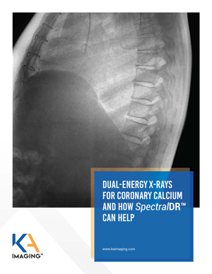 DUAL-ENERGY X-RAYS FOR CORONARY CALCIUM AND HOW 𝘚𝘱𝘦𝘤𝘵𝘳𝘢𝘭𝐃𝐑ᵀᴹ CAN HELP
The world is experiencing an alarming increase in cardiovascular disease. In the US, for example, cardiovascular disease is already America’s costliest disease, but costs are expected to increase to $1.1 trillion USD by 2035.
Coronary calcium provides proof of coronary artery disease; successful detection will improve healthcare for many patients. Traditional Digital Radiography (DR) will usually fail at highlighting coronary calcium due to its poor bone/tissue differentiation. CT is effective; however, it is expensive, and not widely accessible.
Studies show that standard dual-energy scans are better able to detect coronary calcification than standard chest X-rays. However, old approaches of dual-energy have their own limitations – being the overall radiation dose for the procedure one of them. A novel technology, which allows for a single-exposure dual-energy X-ray solution, can overcome these limitations.
Download the white paper to learn more about how SpectralDR™ can help visualize Coronary Calcium and see a real case.
 The mobile X-ray will have increased imaging diagnostic capacity for a variety of patients
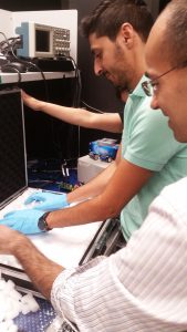


 “‘You are the only girl in the class!’ The sentence I heard a lot and told myself a lot!