
inCiTe 3D X-ray Microscope to support Orthopaedic research at the University of Waterloo
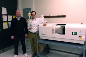 (Waterloo, 25 April, 2024) – X-ray equipment manufacturer KA Imaging has announced the installation of a unit of the inCiTe™ 3D X-ray Microscope at the University of Waterloo. The technology will be used in projects led by Dr. Stewart McLachlin, Assistant Professor in the Mechanical and Mechatronics Engineering department.
The new system is installed in the new Orthopaedic Mechatronics Laboratory, co-led by Dr. McLachlin and Dr. Naveen Chandrashekar. It supports interdisciplinary research interests in studying bone and joint tissue mechanics and interfaces at the microstructural level.
 According to McLachlin, UW is the first research institution in Canada to have access to phase-contrast X-ray µCT imaging for biological tissue characterization.
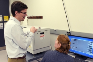
“The inCiTe system fills an important need for our biomechanics research programs. It enables us to see inside the tissues and devices to understand how the microstructure is formed and deformed under varied biological conditions. This is particularly crucial at the interfaces between different materials. The phase-contrast capabilities of the system will help to elucidate these intricate processes,” said Dr. McLachlin.
Musculoskeletal conditions: the leading cause of disabilities in Canada1
Data shows that musculoskeletal (MSK) conditions are the leading cause of disabilities in Canada. Chronic pain – often associated with MSK conditions – has an annual burden of $60 billion, including not only health care costs but also lost wages and taxes2.
The team of researchers at the University of Waterloo is leading innovative biomedical engineering research programs to better understand bone and joint tissue mechanics. New discoveries are expected to lead to improved tools and devices for injury prevention, rehabilitation, and repair.
“The proposed equipment significantly improves image contrast of low-density materials like soft tissues and novel biomaterials, which to date has been a major limitation for characterizing the interface of these materials with traditional µCT imaging.
Further, the near real-time imaging provided by phase-contrast X-ray µCT will enable, for the first time, more accurate and efficient characterization experiments of progressive changes in biological tissue microstructure under different loading conditions,” said Mc. Lachlin.
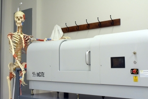
About inCiTe 3D X-ray Microscope
The inCiTe™ 3D X-ray Microscope is the first commercial system that utilizes BrillianSe™, a patented high spatial resolution amorphous selenium (a-Se) detector exclusively developed by KA Imaging Inc.
The high spatial resolution and detection efficiency of the BrillianSe™ X-ray detector enable rapid phase-contrast imaging and conventional micro-CT in a portable benchtop system. Thanks to such phase-contrast technology, the inCiTe™ 3D X-ray Microscope provides superior contrast, and better visualization of low-density materials.
- Kopec JA, Cibere J, Sayre EC, Li LC, Lacaille D, Esdaile JM. Descriptive epidemiology of musculoskeletal disorders in Canada: data from the global burden of disease study. Osteoarthr Cartil. 2019;27:92–S516. doi: 10.1016/j.joca.2019.02.629.
- Parto DN, Wong AY, Macedo L. Prevalence of musculoskeletal disorders and associated risk factors in canadian university students. BMC Musculoskelet Disord. 2023 Jun 19;24(1):501. doi: 10.1186/s12891-023-06630-4. PMID: 37337246; PMCID: PMC10278339.