Entries by Fernanda Fraga

,

KA Imaging to Showcase Growing Family of Reveal Products at RSNA

From detectors to mobile systems, company offerings strengthen its position in the X-ray market


(Waterloo, November 16, 2023) What began as a concept presented at RSNA 2017 has now become a clinically proven solution offered in a variety of products. At the 2023 meeting, Canadian manufacturer KA Imaging is ready to showcase a robust product line that can attend both fixed rooms and mobile systems. “1 exposure, 3 images, 0 motion artifacts”, a summary of the company patented SpectralDR technology, can now be implemented in a variety of environments for better clinical and operational outcomes.


One of the most prominent use cases is the ICU. Medical imaging plays a critical role in monitoring the condition of ICU patients, commonly to check for conditions like pneumonia or pneumothorax or to rule out other potentially serious pulmonary issues along with verifying the tips of catheters or endotracheal tubes. 


Imaging in the ICU can be challenging because of reduced patient mobility, the need for imaging outside regular operating hours, and the need for quick imaging turnaround for bedside decision making.


Generally limited on tissue differentiation, portable chest radiography can be ineffective at accurately spotting complex pulmonary issues and sometimes even to localize the tips of lines and tubes. Other modalities like CT are not portable, bring increased radiation exposure, in addition to risks associated with intra-hospital patient transportation. Furthermore, reimbursement for ICU patients is capitated so an unnecessary CT scan increases cost of care and the financial burden for hospitals.


“Our radiographic spectral images separate materials such as water (i.e., soft tissue, lung lesions etc.) and calcium (i.e. bones, retained foreign objects, in dwelling devices or other calcifications) and are higher contrast thus, easier to read for a variety of clinicians including intensivists, residents and radiologists”, said Dr. Karim S. Karim, CTO of KA Imaging. “Agile decision making is critical in this context. The ability to get increased diagnostic information from a procedure as simple as a chest X-ray in the intensive care unit can simultaneously ease the burden on ICU staff, intensivists, and radiologists,” continued Amol Karnick, President and CEO of the company.


Another condition that can be better seen thanks to dual-energy subtraction are pulmonary nodules. Initial results presented at ECR 2022 reported increased lesion visibility in 43% of the cases thanks to the dual-energy images generated by the company device. The unprecedented capability to offer other views, like laterals or obliques, has also enabled some non-obvious use cases for X-ray, like cardiac calcifications.


“The flexibility of uses for our technology has really made it possible to use dual energy for all general radiography, and it doesn’t have to be limited to a niche like it was before. Single-exposure dual-energy subtraction – our SpectralDR technology inside the Reveal detector – adds confidence without adding radiation or extra work or time”, said Karnick.


KA Imaging is exhibiting at booth 7948 in the North Hall, showcasing the Reveal 35C detector (FDA 510(k) cleared), as well as the company premium mobile system, Reveal Mobi Pro (coming soon). Visitors can also learn more about other solutions, and see some real clinical cases.

Spectral Imaging: The Next Frontier in Medical Diagnostics

Karim S. Karim and Amol Karnick, the visionary co-founders of KA Imaging, recently appeared on SkeletonCrew: The Rad Tech Show to discuss their innovative medical imaging technology. 

In this episode of Skeleton Crew, host Jen Callahan engages in an insightful discussion with Karim S. Karim and Amol Karnick, co-founders of KA Imaging, a Canadian company at the forefront of innovative medical imaging technology. The conversation revolves around the groundbreaking technology developed by them, specifically their portable detector powered by SpectralDR technology. The episode delves into the transformative impact of spectral radiographic imaging on medical diagnostics, patient care, and beyond. Shedding light on the company’s journey, the technology’s capabilities, and its potential applications across various domains.

During the conversation, they talked about the science behind spectral radiographic imaging and the potential applications of this technology in various medical fields. They also highlighted the benefits of  SpectralDR technology and how it can revolutionize medical diagnostics. Their passion for improving patient outcomes through technology is truly inspiring.

watch the episode

,

X-ray Imaging in Agriculture

inCiTe™ 3D X-ray Microscope

BrillianSe X-ray Detector

Applications in Agriculture and Nature


X-ray imaging can play a significant role in agriculture by providing insights into the quality, structure, and composition of crops and agricultural products. Specifically, x-raying can be used for:

  • Quality assessment of seeds and grains
  • Detection of plant diseases and pests
  • Soil structure, compaction and root distribution
  • Post-harvest quality assessment

KA Imaging offers two unique products for X-Raying in Agricultural 

BrillianSe™ is a selenium (a-Se) CMOS direct conversion detector. It provides a unique combination of high spatial resolution and high Detective Quantum Efficiency (DQE) for energies up to 100keV. This combination enables efficient imaging at low flux and high energy, as well as propagation-based (grating-less) phase-contrast enhancement for improved sensitivity when imaging low-density materials.

inCiTe 3D X-Ray Microscope is the first commercial scanner that utilizes BrillianSe™ x-ray detector and is designed with patented propagation-based, phase-contrast imaging to enhance detail of the fine structures that are typically X-ray transparent.


Contact us for more information: sales@kaimaging.com

, ,

KA Imaging Presenting Poster on Innovative Detector Technology at Denver X-ray Conference

WATERLOO, ON. August 2 –  Technical Lead Chris Scott will be presenting the poster on “Direct Conversion 7.8-Micron Pitch Hybrid CMOS X-ray Detector for New Applications in Material” at Denver X-Ray Conference (DXC). The presentation will be on Monday, August 7th, and take place at The Westin Chicago Lombard in Lombard, Illinois, USA from 5 p.m. to 7 p.m.

The BrillianSe X-ray Detector has some novel applications for Dark Field X-ray Microscopy, Bragg Coherent Diffraction Imaging and X-ray Phase Contrast Imaging, as well as applications that require higher X-ray energies and direct X-ray detection.

“Now synchrotron researchers can finally obtain a detector that provides both high spatial resolution and high detection efficiency at higher X-ray energies like 63 keV,” said Karim S. Karim, CTO of KA Imaging.

“We have characterized BrillianSe at x-ray energies up to 63 keV on the beamline 1-BM-B at the Advanced Photon Source,” explained Scott.

The measured responsivity and spatial resolution demonstrate the successful development of a detector technology that can advance research in materials science applications such as high spatial resolution imaging of nanoscale lattice distortions in bulk crystalline materials using Bragg Coherent Diffraction Imaging (BCDI) at energies greater than 50 keV.

The technology can also facilitate the creation of novel synchrotron imaging applications for x-ray energies at or above 20 keV, for example, dark field x-ray microscopy (DFXM) of low diffraction intensity 2D systems such as thin film geometries.

The Denver X-ray Conference is the world’s largest X-ray conference, bringing together experts in X-ray analysis, including both X-ray fluorescence and X-ray diffraction, to discuss the latest advancements in the field. This year, the event will be held in Chicago from August 7 to 11.

In addition to the poster presentation, KA Imaging will participate in the technical exhibition at booth #18.

About KA Imaging

A spin-off from the University of Waterloo, KA Imaging is a company that specializes in developing innovative X-ray imaging technologies and systems, providing solutions to the medical, veterinary, and non-destructive test industrial markets.

For further information: Media Inquiries, please contact: media@kaimaging.com

,

KA Imaging To Launch Premium Dual-Energy Mobile System at AHRA 2023

WATERLOO, July 6, 2023 – X-ray manufacturer KA Imaging is unveiling its Reveal Mobi Pro dual-energy mobile X-ray system. The product, which is a collaboration with UMG/Del Medical, will be showcased at the upcoming AHRA Annual Meeting.

“This partnership with Del Medical comes at an excellent time,” said Amol Karnick, President and CEO of KA Imaging. “After the pandemic, mobile imaging has become more prominent in many healthcare facilities, so offering a solution to improve the visualization of bones and tissues at the bedside is essential,” said Karnick.

“We are excited to support KA Imaging and their enhanced detector technology for the conventional and mobile radiographic markets,” said Marc Lorenzo, Executive Vice President at Del Medical.

The Reveal Mobi Pro integrates KA Imaging’s Reveal™ 35C detector with SpectralDR™ technology into a complete mobile X-ray solution. Reveal’s ability to simultaneously acquire conventional and dual-energy images with a single exposure at the bedside improves hospital and patient outcomes and protects revenue by reducing outflows.

The Reveal 35C detector mimics the workflow, dose and techniques of state-of-the-art mobile DR X-ray. With the Reveal Mobi Pro, it is offered in a fully integrated mobile system.

Last November, KA Imaging unveiled its first integrated mobile dual-energy X-ray system, Reveal Mobi Lite. The main difference between the Lite and Pro versions are around some advanced features, only available in the Pro system. The most noteworthy feature of the Reveal Mobi Pro is the telescopic arm. In addition to the collapsible column design, the system has a high-frequency 40KW generator, integrated DR Workstation, 8-hour battery life, and motor-assisted movement.

The AHRA Annual Meeting will be held from July 9-12 at Indiana Convention Center. KA Imaging has booth #329.

The Reveal 35C detector is FDA 510(k) cleared. The Reveal Mobi Pro is coming soon and is not available for sale.

About KA Imaging 

A spin-off from the University of Waterloo, KA Imaging specializes in developing innovative X-ray imaging technologies and systems, providing solutions to the medical, veterinary, and non-destructive test industrial markets.

About UMG/DEL MEDICAL

UMG/DEL MEDICAL is a leading source of innovative radiographic products, with 90 years in medical imaging. We are primarily engaged in the design, manufacturing and distribution of high performance digital and analog medical imaging systems, sold and serviced globally by factory trained and authorized medical equipment sales and service professionals. Our extensive product portfolio of radiographic components, systems and accessories is designed to support hospitals, imaging centers and clinics by tailoring systems to accommodate each facility’s specific requirements and budget. UMG/DEL MEDICAL’s products are manufactured, pre-staged and tested in our Bloomingdale, IL and Harrison, NY facilities.


For further information
Media Relations
media@kaimaging.com 
T: 226.215.9897

,

Health Check Bus: Lung Screening Project in Taiwan Reaches 8,000 Patient Mark

Mobile Unit is equipped with KA Imaging’s Reveal 35C


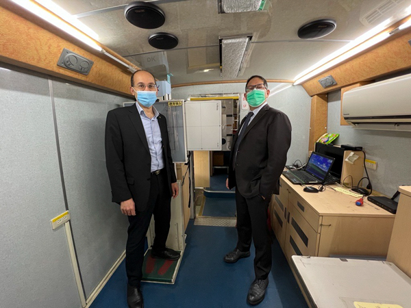
More than 8,000 patients have already been imaged by the Health Check Bus in Taiwan. The project started in July 2022 in Yunjianan and Penghu, an area comprising the county of Yunlin, Chiayi, Penghu and Tainan city. It is a cooperation between InnoCare Optoelectronics and the College of Medicine at National Cheng Kung University (NCKU) and focuses on mobile X-ray lung screening.

The Health Check Bus is equipped with KA Imaging’s Reveal 35C. Reveal™ 35C is a single exposure, portable, digital dual-energy subtraction (DES) X-ray detector. Powered by SpectralDR™ technology, it uses the same radiation dose as a chest X-ray to create three different images without motion artifacts. In medical applications, this means a regular DR, plus a soft tissue and bone images.

Although Low Dose Computed Tomography (LDCT) is the current gold standard for lung cancer screening, it still has a few limitations. To be imaged, patients must visit the hospital; however, equipment distribution and availability is uneven. Therefore, having a high-volume screening tool that can be taken to patients and offer better insights than traditional X-ray can be a game changer.

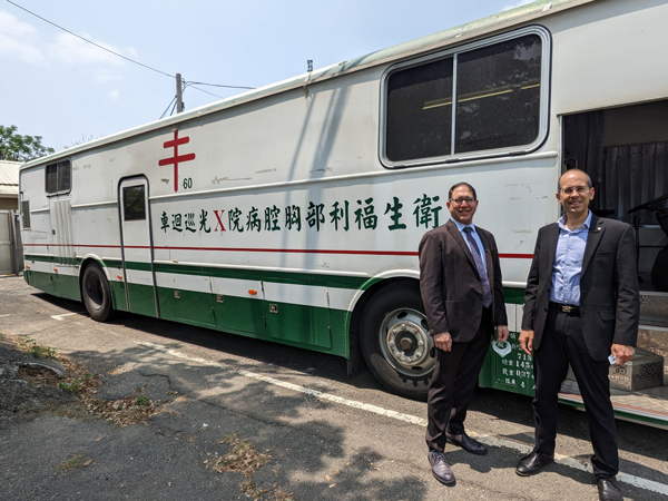
The project is the largest epidemiological screening of lung cancer in Taiwan. Its results will be used as a reference for future changes in lung cancer prevention and control policies. It is expected to image 10,000 people.

“Reveal 35C has already been clinically proven to better detect lung lesions”, said Amol Karnick, President and CEO of KA Imaging. “Our studies show that radiographic spectral images can play an important role in earlier detection, and we’re excited to see how our technology will improve outcomes in such a high-volume project” he continued.

James Yang, chairman of InnoCare Optoelectronics, pointed out that “the additional soft tissue and bone images can help doctors diagnose a number of chest diseases early, and could be used further in any health check up projects, including the ones targeting remote rural areas, greatly benefiting patients and medical institutions.” According to InnoCare, access to lung screening tools in remote areas is less than 30% in Taiwan.  

, ,

KA Imaging installs inCiTe 3D X-ray Microscope and partners with the University of Greenwich

Technology will support research in biological tissues, biomaterials and other engineering materials.

(Waterloo, 24 April 2023) – X-ray equipment manufacturer KA Imaging has announced a new install of the inCiTe™ 3D X-ray Microscope as part of a strategic partnership with the University of Greenwich, United Kingdom. The technology, first of its kind available in UK/Europe, will be used in research and knowledge transfer projects led by Professor Gianluca Tozzi.

The inCiTe™ 3D X-ray Microscope is the first commercial system that utilizes BrillianSe™, a patented high spatial resolution amorphous selenium (a-Se) detector exclusively developed by KA Imaging Inc.

The high spatial resolution and detection efficiency of the BrillianSe™ X-ray detector enable rapid phase-con­trast imaging and conventional micro-CT in a portable benchtop system. Thanks to such phase-contrast technology, the inCiTe™ 3D X-ray Microscope provides superior contrast, and better visualization of low-density materials.

The new inCiTe™ unit will support joint research efforts aimed at further advancing fast phase-contrast tomography of soft tissues and biomaterials in the laboratory.

“inCiTe’s phase contrast technology is particularly beneficial for objects with low conventional absorption contrast, such as the soft biological tissues studied by Professor Tozzi,” explained Chris Scott, Technical Lead at KA Imaging. “We had very promising initial results with some samples we processed in inCiTe, where bone and soft tissue can be better visualized thanks to phase-contrast,” he continued.

Professor Gianluca Tozzi is Professor of Industrial Engineering at the Faculty of Engineering and Science, University of Greenwich, United Kingdom. His research is devoted to the understanding of structure/function of biological tissues and biomaterials using experimental techniques such as such as digital volume correlation (DVC) based on X-ray computed tomography, aimed at informing product development for industry as well as clinical practice for traumatic and pathological conditions. “I am very excited by the unique capability of the inCiTe 3D X-ray Microscope and potential for further technological development of phase-contrast tomography, within this partnership with KA Imaging,” said Professor Tozzi. “,” said the Professor.

,

Soft Tissue Image by KA Imaging’s Reveal 35C Undercovers Mass Behind In-Dwelling Device

WATERLOO, ON– Manufacturer KA Imaging announced a study examining “single-exposure dual-energy radiography for the subtraction of in-dwelling devices in the soft tissue image” that made use of the Reveal 35C X-ray detector, powered by SpectralDR™ technology.

Using the Reveal 35C detector, the purpose of the study was to determine the clinical value of removal of in-dwelling devices in the dual-energy soft tissue X-ray image. According to the study, the spectral radiographic images revealed a suspicious soft tissue mass in one of the cases by subtraction of an overlapping pacemaker in the soft tissue image1.

Images were taken from 23 patients having at least one in-dwelling device, and the patients were all part of an on-going 600 patient study using the Reveal 35C detector at Taipei Medical University Hospital in Taiwan.

“The fact that the soft tissue image was able to find the mass, not seen in the conventional DR image, overlapping a pacemaker is quite promising and indicates that SpectralDRTM technology can efficiently subtract, in addition to bone calcium, higher atomic number materials such as metals associated with the pacemaker,” said Dr. Karim S. Karim, CTO of KA Imaging. “Spectral radiographic images generated by SpectralDRTM can play an important role in early disease detection for better outcomes,” says Amol Karnick, President and CEO.

The study will be presented at this year’s European Congress of Radiology (ECR). In addition to the e-poster at the scientific congress, KA Imaging is part of the technical exhibition at Expo X4, booth 437.

ECR is one of the largest medical meetings in Europe and the second-largest radiological meeting in the world.

SpectralDR™ technology enables dual-energy subtraction, providing bone and tissue differentiation with a single standard X-ray exposure. It acquires three images simultaneously (DR, bone and soft tissue dual-energy X-ray images). The technology mimics the workflow, dose and techniques of state-of-the-art mobile DR X-ray detectors.

About KA Imaging

A spin-off from the University of Waterloo, KA Imaging specializes in developing innovative X-ray imaging technologies and systems, providing solutions to the medical, veterinary, and non-destructive test industrial markets.


  1. P-C. Chou, K. S. Karim. “Single-exposure dual-energy radiography for the subtraction of in-dwelling devices in the soft tissue image”. European Congress of Radiology-ECR 2023, 2023.


Contacts:


Fernanda Fraga 

Media Relations
ffraga@kaimaging.com 
T: 226.215.9897

,

KA Imaging Presents Technology for Better Material Separation At SPIE 2023

KA Imaging CTO Dr. Karim S. Karim is attending the SPIE International Medical Imaging Symposium, being held in San Diego from February 19 to 23, where presented the study “Multimodal single-shot x-ray phase-contrast imaging using a high-resolution micropillar-based grating”. The study, a part of engineer Abdollah Pil-Ali’s PhD dissertation, was co-authored by Abdollah Pil-Ali, Sahar Adnani and Dr. Karim from the University of Waterloo, as well as Serena Qinyun Z. Shi and Professor Dr. Peter B. Noël from the University of Pennsylvania.

In this work, the authors present a novel approach to build a large area high resolution X-ray grating for emerging phase contrast X-ray imaging applications.  Like optical light undergoing refraction when passing through a prism, X-rays also refract when passing through objects. However, X-ray refraction is very minor akin to seeing a football stadium from the moon and usually requires a synchrotron source. An X-ray grating helps amplify the refracted signal and thus allows generation of three images with one single exposure: attenuation, phase and scatter in a compact laboratory setup. The simultaneous presence of attenuation and scatter images can help with both material composition and material identification e.g., telling the difference between fine and coarse powders or even the difference between aluminum and steel.

SPIE is the international society for optics and photonics. The SPIE Medical Imaging meeting, to be held in San Diego, offers an opportunity to hear the latest advances in image processing, physics, computer-aided diagnosis, perception, image-guided procedures, biomedical applications, ultrasound, informatics, radiology, and digital and computational pathology.