
Micro-computed tomography (micro-CT) has been a key imaging technology for non-destructive testing (NDT), materials analysis, and biomedical research for decades. However, as modern materials become increasingly complex—with multi-material assemblies, fine microstructures, and low-density components—the limitations of conventional absorption-based micro-CT become more evident.
To address these evolving challenges, micro-CT technology has expanded beyond simple absorption contrast, integrating new contrast mechanisms like phase contrast and spectral imaging. KA Imaging’s inCiTe™ 3D X-ray Microscope platform leverages both of these complementary approaches, enabled by advanced detector technologies: BrillianSe™ and Reveal™.
The Core Limitation: Absorption Contrast Alone Isn’t Enough
Traditional micro-CT relies on X-ray attenuation — how much energy is absorbed by materials as X-rays pass through. High-density materials absorb more, while low-density materials absorb less. However, when different materials have similar attenuation profiles, or when critical features are extremely small, absorption contrast alone is often insufficient.
This creates challenges across industries:
- Semiconductors and electronics: detecting micro-voids, cracks, and thin interconnect layers
- Composites and additive manufacturing: resolving fiber alignment, porosity, and delamination
- Biomedical research: visualizing soft tissues and implants without destructive sample prep
- Multi-material assemblies: differentiating between polymers, metals, and layered structures
Simply increasing resolution or dose is not always feasible, especially for delicate samples or high-throughput environments. New contrast mechanisms are required to overcome these limitations.
Multiple Contrast Mechanisms: Two Paths for Evolving Imaging Needs
Path 1: High-Resolution Phase Contrast Imaging — Visualizing Fine Structures and Low-Density Materials
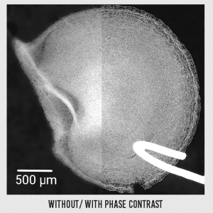 Phase contrast imaging captures not only how much X-rays are absorbed, but also how their wavefront phase shifts as they pass through different materials. This dramatically improves contrast for low-density and weakly absorbing materials that are difficult to image with absorption contrast alone.
Both configurations of the inCiTe™ platform utilize propagation-based phase contrast — a grating-less method that captures interference patterns as X-rays propagate beyond the sample, enhancing boundary definition and internal detail.
However, when paired with KA Imaging’s BrillianSe™ direct conversion detector, phase contrast imaging reaches its full potential:
- 8 µm pixel size enables submicron voxel resolution (down to 0.8 µm)
- High Detective Quantum Efficiency (DQE) at both low and high energies
- Direct a-Se conversion eliminates optical blurring from scintillators
- Exceptional boundary sharpness and small feature detectability
This configuration is particularly powerful for:
- Polymers and composites
- Thin films and coatings
- Soft biological tissues
- Microelectronics and interconnect structures
With BrillianSe™, inCiTe™ 3D X-ray Microscope delivers highly detailed, submicron-resolution phase contrast imaging without the need for staining, slicing, or destructive preparation.
Path 2: Spectral Imaging with Phase Contrast Benefits — Differentiating Materials by Composition
While phase contrast reveals fine structure, it does not inherently differentiate materials based on their elemental composition. This is where spectral (multi-energy) CT becomes essential.
Spectral CT measures how materials absorb X-rays at multiple energies, enabling compositional analysis by separating materials with overlapping density but differing atomic numbers.
Applications include:
- Separating metals from polymers in over-molded electronics
- Identifying foreign inclusions in aerospace and composite parts
- Analyzing electrode materials in batteries
- Layer differentiation in multi-material assemblies
In this configuration, KA Imaging’s Reveal™ detector plays a central role. With its proprietary triple-layer design, Reveal captures multiple energy bands simultaneously in a single exposure, eliminating filter swapping and reducing motion artifacts. At the same time, the propagation-based geometry allows Reveal to also benefit from phase contrast enhancement — especially at higher magnifications or when imaging low-density structures within large parts.
While spatial resolution is lower (140 µm pixel size), this configuration offers an ideal balance for large samples where composition analysis and multi-material separation are critical. The inCiTe™ 2.0 platform integrates these spectral capabilities while still leveraging phase-based edge enhancement.
Detector Technology Defines What’s Possible
| Feature | ||
|---|---|---|
| Detector Type | Direct Conversion (a-Se / CMOS) | Triple-layer Indirect Conversion (CsI-based) |
| Key Benefit | Submicron resolution and fine structural detail | Multi-energy material separation |
| Resolution | 8 µm pixel / 0.8 µm voxel | 140 µm pixel |
| Phase Contrast Capability | Optimized for highest phase sensitivity | Phase enhancement with spectral contrast |
| Ideal Use Cases | Composites, semiconductors, biomedical tissues | Multi-material assemblies, aerospace, electronics, batteries |
inCiTe™ Platform: Modular Imaging for Complex Materials
KA Imaging’s inCiTe™ platform provides laboratories and manufacturers with modular tools to meet a wide spectrum of imaging challenges:
BrillianSe configuration (inCiTe 3D X-ray Microscope):
- High-resolution phase contrast (submicron detail)
- Low-dose operation with high DQE
- Ideal for fine structure and soft material visualization
Reveal configuration (inCiTe 2.0 3D X-ray Microscope):
- Simultaneous multi-energy spectral acquisition
- Efficient material composition separation
- Phase-enhanced structural contrast for larger samples
This flexible architecture allows users to select the optimal configuration for their materials, sample sizes, and inspection needs — all within a compact, benchtop 3D X-ray microscope system.
Conclusion: Expanding What Micro-CT Can Reveal
The increasing complexity of modern materials requires imaging technologies that go far beyond conventional CT. Both phase contrast and spectral imaging have become essential tools for accurately characterizing fine structures and material compositions across industries.
By offering both contrast mechanisms within the inCiTe™ platform — powered by BrillianSe and Reveal detectors — KA Imaging enables researchers and engineers to fully resolve today’s most advanced materials.
inCiTe™: Spark New Possibilities. Imaging breakthroughs start here.