
 In critical care, clarity isn’t a luxury — it’s a necessity.
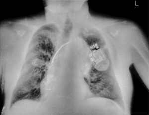
Consider the following case: a mechanically ventilated ICU patient shows signs of respiratory distress. A portable chest X-ray is ordered. On the standard digital radiograph, the region of interest in the right lung appears ambiguous. Overlapping ribs and an ECG clip obscure the view — making it difficult to determine whether a consolidative process is evolving. There’s no time to reposition the patient. CT is not an option.
 What if you could remove the visual noise?
With SpectralDR® technology in the Reveal™ 35C detector, that same exposure produces a soft-tissue-only image — subtracting the bone and medical hardware. The result: a clearly defined area of consolidation in the right lung, enabling a more confident, real-time clinical decision.
This is the power of dual-energy imaging brought to the bedside. And in high-acuity environments like the emergency department (ED) and intensive care unit (ICU), every image must pull its weight.
When Standard Imaging Falls Short
Conventional X-ray remains the workhorse of hospital imaging — but in high-stakes environments, its limitations are exposed:
- Overlapping anatomy obscures key structures
- Diagnostic ambiguity increases reliance on follow-up CTs
- Portable imaging often suffers from motion artifacts and poor positioning
In unstable patients or crowded units, waiting for cross-sectional imaging isn’t always possible. Dual-energy imaging offers a practical, portable upgrade that meets this moment — with no change in workflow.
Mounting Pressure in Critical Care
Canada: Overcrowding and Capacity Strain
- As of 2022–23, Canadian hospitals reported an average acute care occupancy rate exceeding 90%, leaving minimal surge capacity1.
- “Exit block” delays persist—85% of ED directors cite a lack of admitting beds as a key bottleneck2.
United States: Resource Constraints and Throughput Challenges
- U.S. ICUs average 68–75% occupancy, with projected increases to 85% by 20323, 4.
- The U.S. sees 155 million ED visits annually; 3.1 million lead to ICU admission5.
- 50% of EDs are routinely overcrowded, and ambulance diversions occur in one-third of hospitals6.
- Up to 10% of patients leave before being seen due to wait times7.
In both systems, imaging delays can compound treatment delays. Rapid, reliable bedside tools are no longer optional — they’re essential.
Reveal 35C: Diagnostic Clarity at the Point of Care
Reveal™ Mobi Pro, powered by the Reveal 35C detector with SpectralDR® technology, delivers three co-registered images from a single exposure:
- Standard radiograph (SE)
- Bone-only view
- Soft tissue-only view
These differentiated views enhance visualization of pulmonary and mediastinal abnormalities while reducing the need for follow-up imaging. And unlike traditional dual-shot methods, SpectralDR® is fully compatible with portable and upright use, requiring no workflow changes or additional radiation.
Proven Performance in the Field
- 33% more pneumonia cases detected compared to conventional X-ray8
- 43% improvement in lesion visibility when dual-energy views were included9
- Up to 37% fewer follow-up CT scans needed in ICU workflows10
- 57% increase in diagnostic confidence, with no added reading time11
Seeing the Unseen: Foreign Object Detection
Portable chest X-rays account for 40% of global imaging, but standard images often miss small or obscured foreign objects:
- Over 50% of tracheal and 25% of bronchial foreign bodies are missed on initial radiographs12
- Subtle objects like metallic foils or embedded glass may go undetected, especially in pediatric or trauma patients
Dual-energy imaging improves contrast separation, enhancing visibility of devices and hardware even in compromised imaging conditions.