Emergency departments rely on imaging to evaluate injuries, guide treatment decisions, and support patient stabilization.
Portable chest X-ray is frequently used in trauma environments because it allows clinicians to image patients immediately in the trauma bay without transporting unstable patients.
However, overlapping anatomical structures can obscure clinically relevant findings in conventional radiographs.
Spectral X-ray imaging expands the information available during emergency radiography. KA Imaging’s Reveal™ platform uses SpectralDR® technology to generate multiple image types from a single exposure—including conventional, bone, and soft-tissue images—while maintaining the same workflow and radiation exposure as standard X-ray.
Solutions for Emergency & Trauma Imaging
Reveal™ spectral imaging solutions can be used in emergency departments and trauma centers.
Emergency imaging plays a central role in evaluating traumatic injuries and acute medical conditions.
In trauma and emergency environments, clinicians often need imaging to assess:
pneumothorax or other life-threatening chest injuries
rib fractures and skeletal trauma
lung disease or infection
device placement following emergency procedures
Spectral imaging can enhance visualization of anatomical structures and pathology by separating bone and soft tissue information into multiple images generated from a single exposure.
Clinical studies using the Reveal 35C suggest:
33%
more pneumonia cases found compared to X-ray thanks to dual-energy images⁴
43%
more lesion visibility when dual-energy images were included³
Better
visibility of lines and tube tips without extra reading times⁵
This additional diagnostic information may help clinicians identify abnormalities that may be less visible in conventional radiographs.
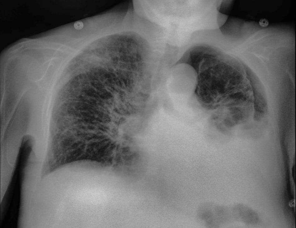
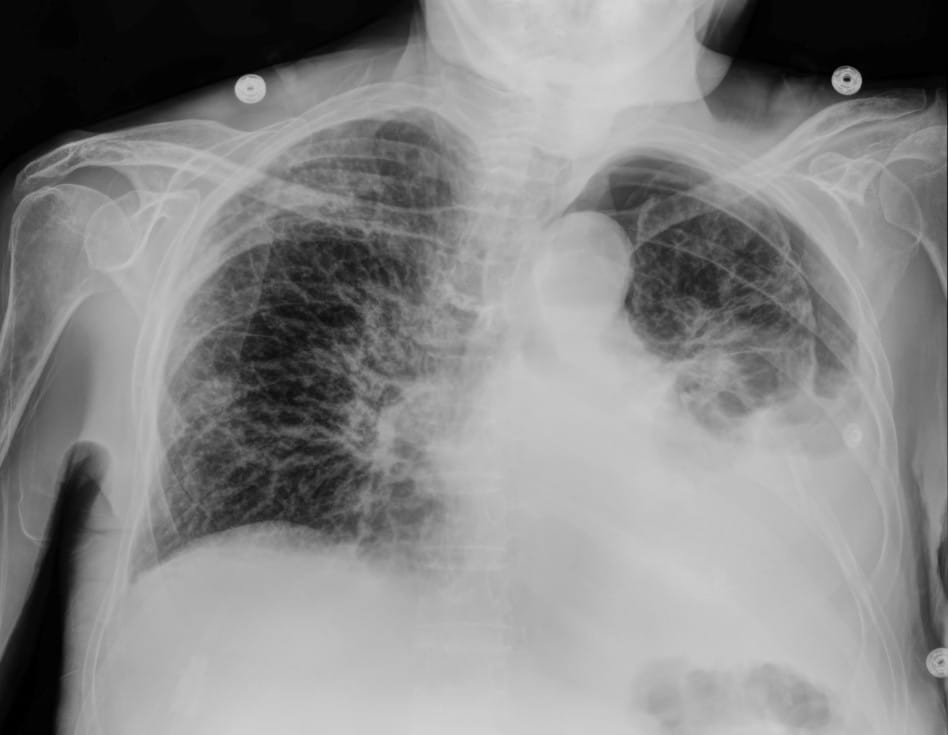
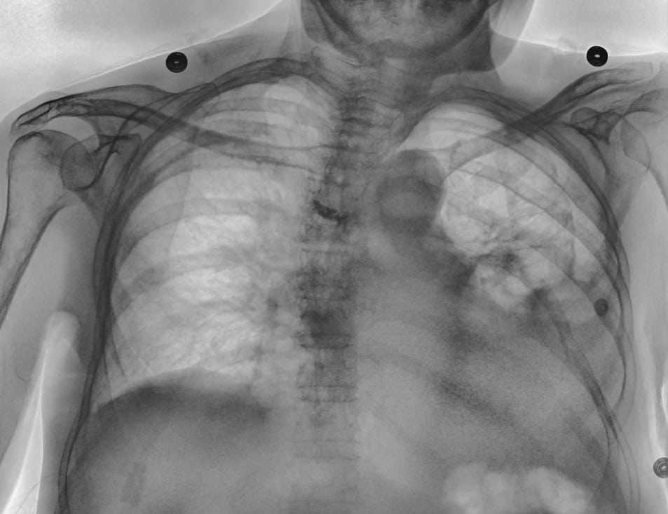
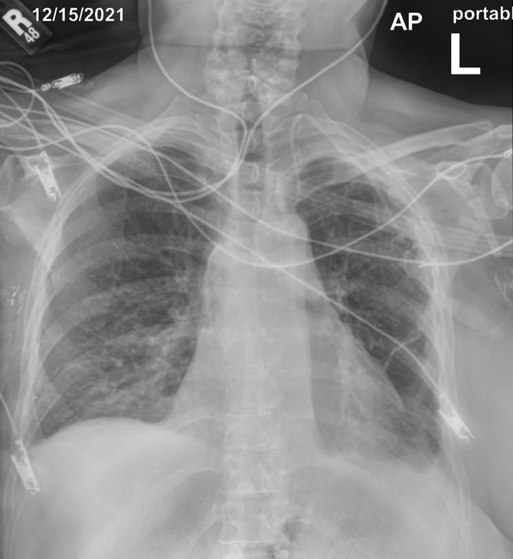
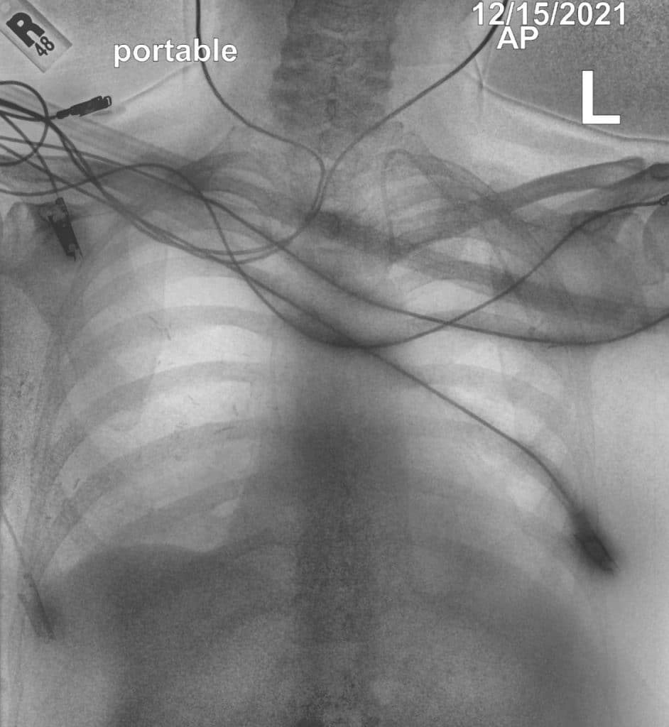
In this case, the conventional DR image shows subtle findings. The spectral soft-tissue image reduces overlying bone, making the pneumothorax more conspicuous. The corresponding bone-selective image highlights a rib fracture, a potential underlying cause of the pneumothorax.
The DE Bone image picks up and delineates the surgical clips in the patient’s right thorax, as well as the end of the tortuous PICC line.
Spectral Imaging at the Point of Care
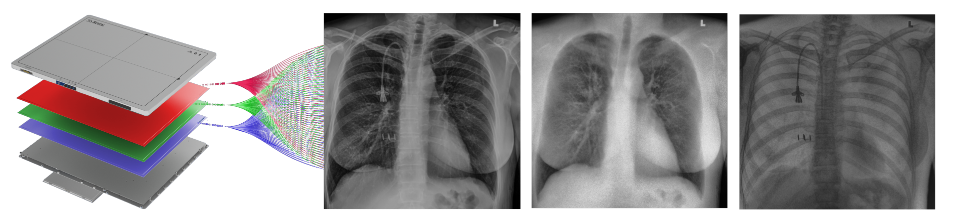
Reveal™ 35C detectors use SpectralDR® technology to acquire multiple spectral images from a single X-ray exposure.
Each exposure produces:
a conventional digital radiography image
a bone image
a soft-tissue image
These images allow clinicians to separate overlapping anatomical structures and visualize areas that may be less visible in conventional radiographs.
Unlike traditional dual-energy imaging techniques that require multiple exposures, SpectralDR captures these images simultaneously with a single exposure, eliminating motion artifacts and maintaining the same clinical workflow as standard portable radiography.
Key Clinical Scenarios in Emergency & Trauma Imaging
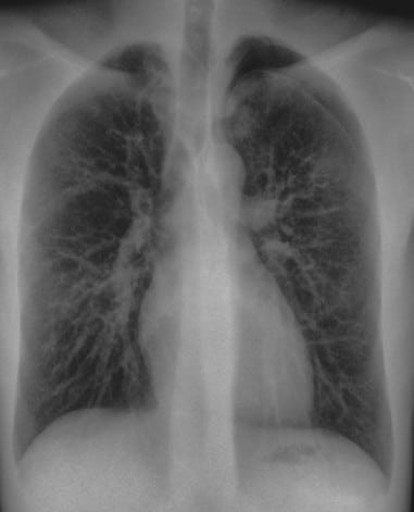
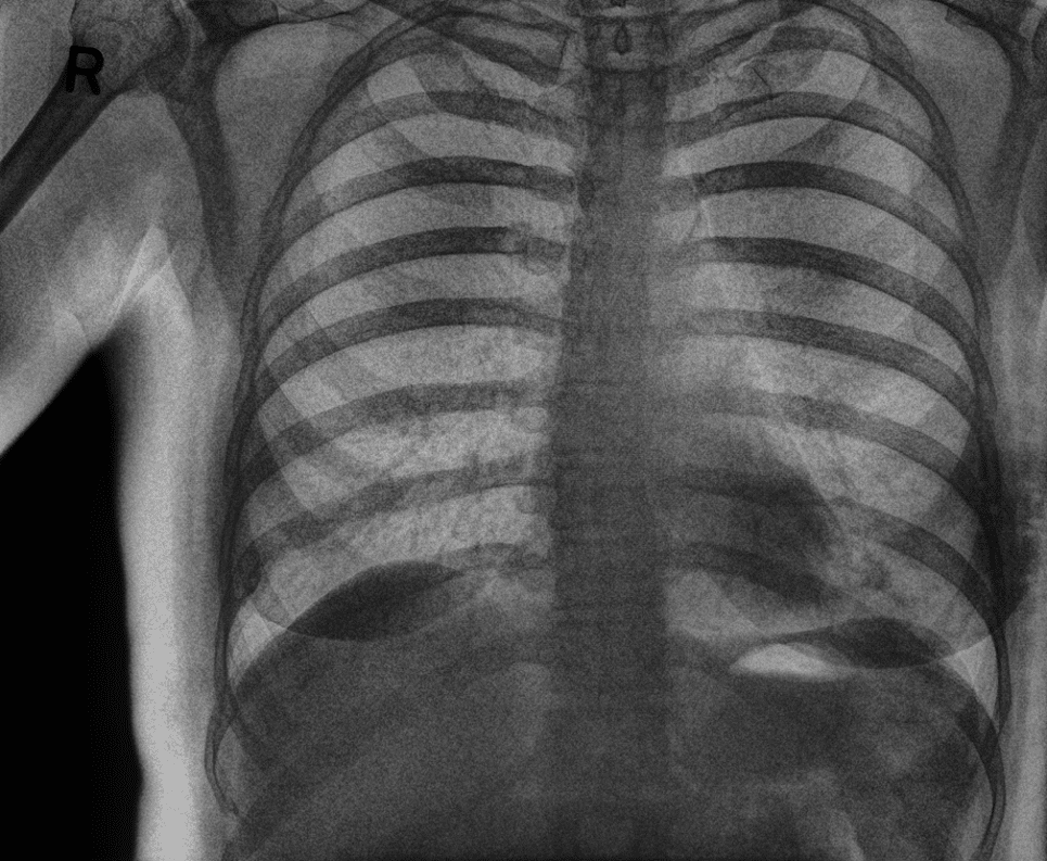
Pneumothorax Evaluation
Soft-tissue images reduce rib overlap and may improve visualization of pneumothorax in trauma patients.
This 26-year-old male presented to the ER at 3am, with difficulty breathing and chest pain. The Reveal 35C Spectral DR detector was used to generate all 3 images. A left apical pneumothorax can be visualized, and is best seen in the Soft Tissue image.
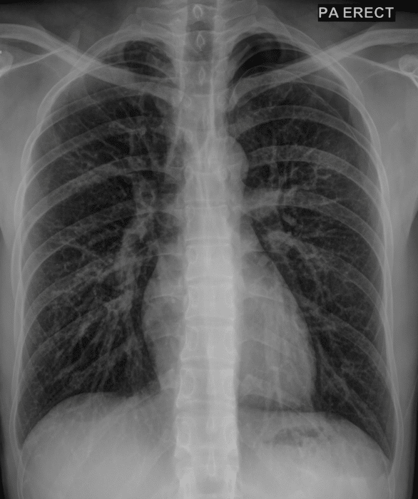
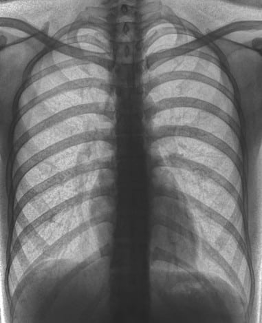
Rib and Skeletal Trauma
Bone images can highlight skeletal structures and fractures that may be difficult to identify in conventional radiographs.
In its ability to remove superimposing soft tissue from images, viewers can better visualize the bony anatomy in greater detail and clarity. In this case, the reader noticed a calloused rib fracture that became visible on the right side.
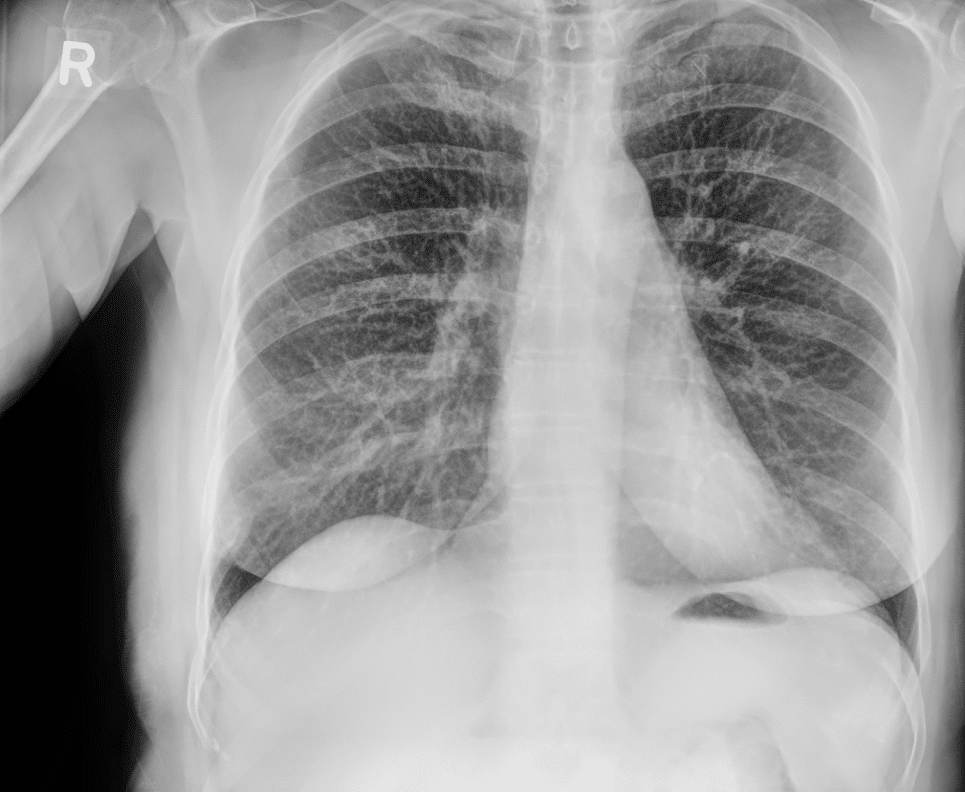
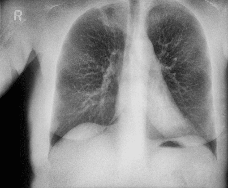
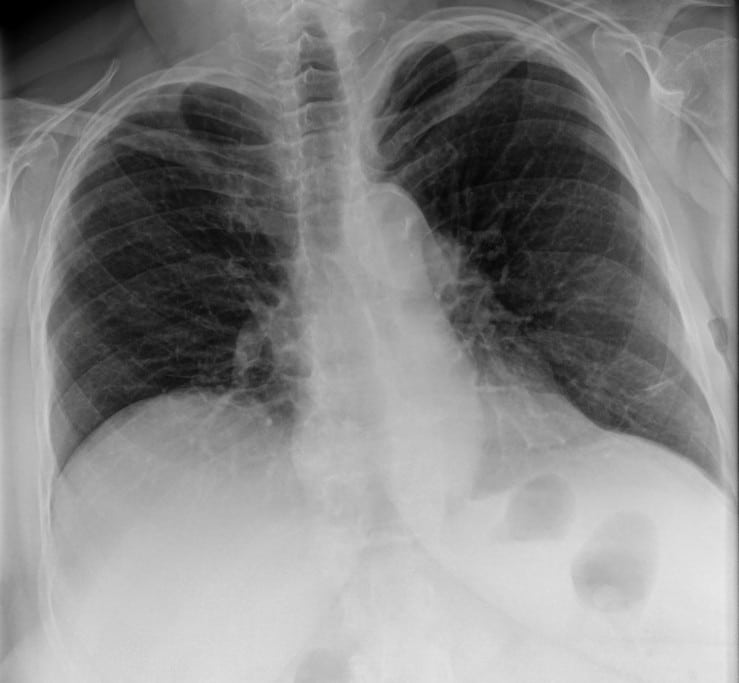
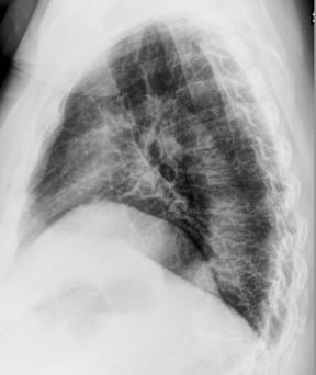
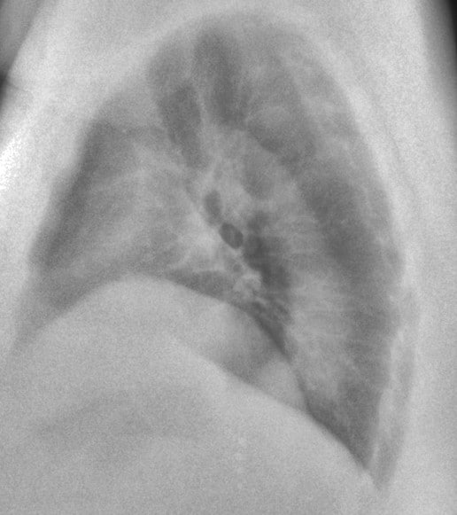
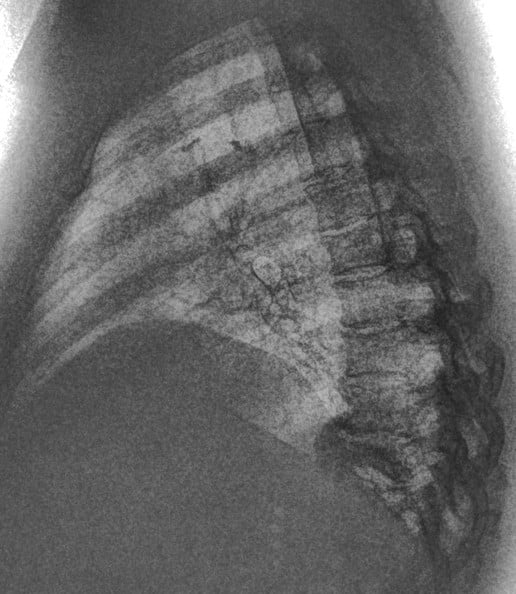
Lung Injury and Pulmonary Abnormalities
Spectral imaging can improve visualization of lung opacities by separating bone structures from soft tissue.
This 67-year old female patient presented to the ER with shortness of breath, in conjunction with chronic pain in her posterior thoracic region upon taking a large breath in. The reading radiologist noticed a suspicious mass-like opacity in the Lateral view. The radiologist stated that, thanks to the DE images, they could they tell the opacity wasn’t a lung mass, but instead far less concerning bony outcroppings from the thoracic vertebrae.