
Cardiovascular Imaging
Visualizing Coronary Artery Calcium in Routine Chest Radiography

Coronary artery calcium (CAC) is a well-established imaging marker associated with coronary artery disease and cardiovascular risk. Traditionally, CAC is evaluated using cardiac CT imaging.
Recent advances in spectral X-ray technology suggest that the clinical value of chest radiography can be expanded. Spectral imaging may enable visualization of coronary calcium during routine chest X-ray exams.
KA Imaging’s Reveal™ platform introduces a new approach to chest imaging using SpectralDR® technology. This single-exposure spectral X-ray detector generates multiple image types — including conventional, bone, and soft-tissue images — from a single exposure while maintaining the same workflow and radiation exposure as standard radiography.
These additional images may improve visualization of calcified structures within the thorax, including coronary artery calcium.
Clinical Evidence
Clinical studies have evaluated the ability of spectral chest radiography to visualize coronary artery calcium.
Clinical evaluations comparing spectral radiography images with traditional X-ray have suggested:
- Opportunistic visualization of CAC and valve/vascular calcium with up to a 61.8% increase in sensitivity compared to standard X-ray, and an AUC of 91% relative to low dose CT¹⁰
- Higher sensitivity and reader agreement for detecting coronary artery calcification¹¹, ¹².
- Dual-energy chest X-ray showed improved performance over standard X-ray, with results approaching those of non-gated CT for detecting coronary calcification¹¹, ¹²
Importantly, spectral imaging generates these additional images using a single exposure, maintaining the same workflow as conventional radiography.
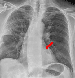
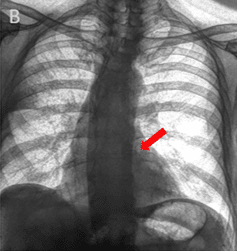
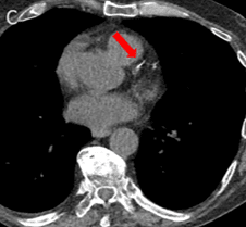
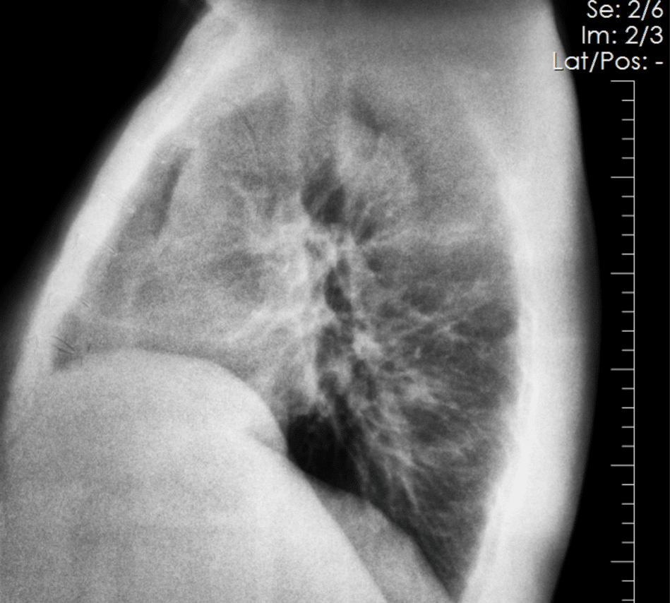
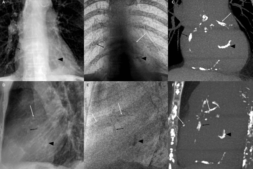
Coronary artery calcium becomes visible in the SpectralDR bone image and is confirmed on CT.
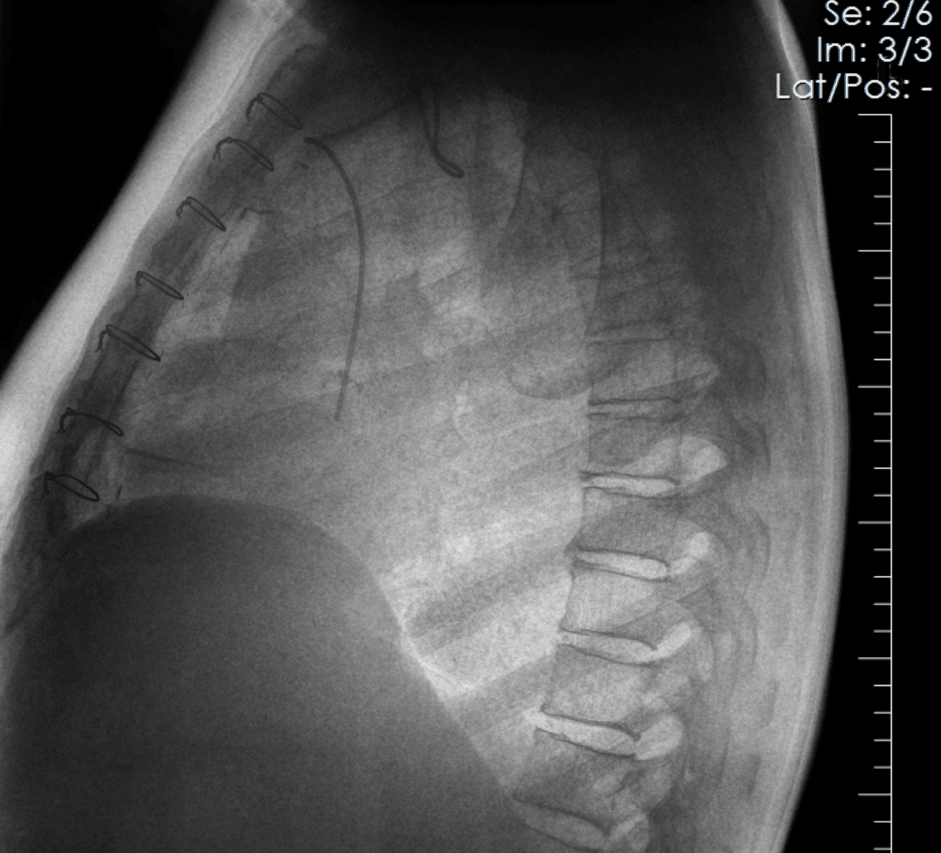
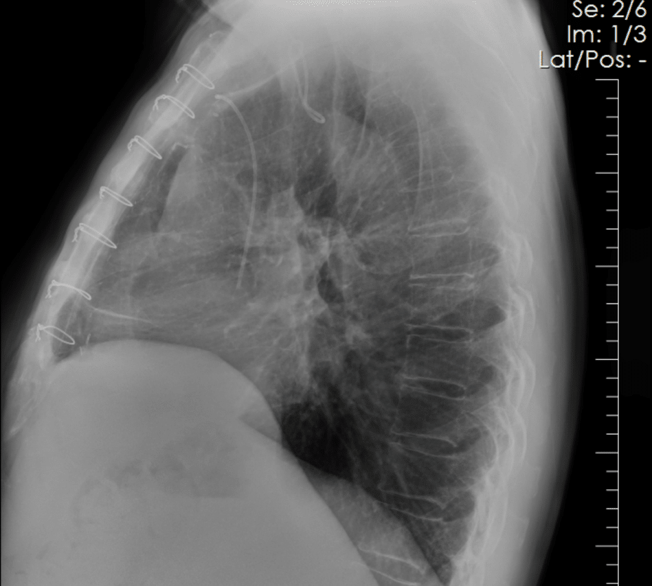
SpectralDR bone images emphasize skeletal structures, allowing clearer visualization of the fracture compared with the conventional radiograph.
Solutions for Cardiovascular Imaging
Reveal™ spectral imaging solutions can be used during routine chest radiography.

Spectral Imaging for Better Visualization of Coronary Calcium
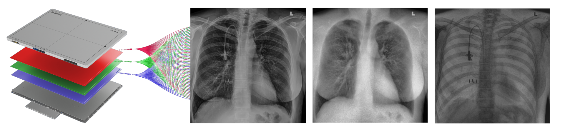
Reveal™ detectors generate three images from a single X-ray exposure:
• conventional radiograph
• bone image
• soft-tissue image
These images allow clinicians to separate overlapping structures and evaluate thoracic anatomy in different ways.
Soft-tissue images can reduce the visual impact of rib structures, while bone images highlight calcified structures within the thorax.
This separation may improve visualization of coronary artery calcium within routine chest radiography exams.
Unlike traditional dual-energy imaging techniques that require multiple exposures, SpectralDR captures these images simultaneously with a single exposure, eliminating motion artifacts and maintaining the same workflow as standard radiography.
Why Spectral X-ray Matters
In conventional chest radiographs, overlapping anatomical structures can obscure small calcifications within the thorax.
Spectral X-ray imaging separates bone and soft-tissue information into multiple image outputs from a single exposure.
This separation can reduce overlapping structures and improve visualization of calcified anatomical features.
Because these images are generated during a routine chest radiography exam, spectral imaging may provide additional imaging context without requiring changes to workflow or radiation dose.
Clinical Context
Chest radiographs are among the most frequently performed imaging exams in healthcare systems worldwide.
Because of their widespread use, chest radiographs may provide opportunities for clinicians to observe imaging features beyond the original indication for the exam.
Spectral imaging expands the information available within routine radiography by providing additional image types that may reveal anatomical features not easily visible in conventional radiographs.
This may contribute additional context when evaluating thoracic anatomy in clinical practice.
Related Applications
Emergency & Trauma Imaging
Chest Imaging
Critical Care & Bedside Imaging

Expand Cardiovascular Insight in Chest Radiography
Learn how spectral X-ray imaging can provide additional imaging information during routine chest radiography.
Latest blogs
Imaging plays an important role in monitoring patient health and supporting clinical decision-making in intensive…
Tuberculosis remains one of the world’s deadliest infectious diseases. Currently, the critical challenge has shifted…
In 2015, KA Imaging was founded with one major goal: to redefine what X-ray technology…