
Biomedical research often requires scientists to visualize structures with vastly different X-ray attenuation properties within the same specimen. Whether studying musculoskeletal disease, tissue-engineered scaffolds, orthopedic implants, or cancer progression, researchers need to examine mineralized tissues alongside soft tissues without sacrificing image quality.
Conventional micro-computed tomography (micro-CT) excels at visualizing dense structures such as bone, but revealing soft tissues within the same scan remains challenging. At KA Imaging, we’ve spent years studying this problem because we believe laboratory X-ray imaging should provide researchers with a more complete understanding of complex biological specimens—not force them to choose between imaging one tissue type or another.
Advances in phase-contrast and spectral imaging are expanding what researchers can see within a single dataset, providing additional information beyond conventional absorption-based imaging and opening new possibilities for biomedical research.
Why Imaging Bone and Soft Tissue Together Is Challenging
The difficulty begins with the way different biological tissues interact with X-rays.
Bone is highly mineralized and absorbs X-rays efficiently, producing strong image contrast in conventional X-ray and micro-CT systems. Soft tissues—including cartilage, ligaments, tendons, muscle, and connective tissue—attenuate X-rays much less, resulting in comparatively weak contrast.
For many biomedical applications, these tissues must be evaluated together. Researchers investigating joint degeneration need to visualize cartilage adjacent to bone. Implant developers need to study both the implant interface and the surrounding biological response. Tissue engineering researchers must evaluate scaffolds while also examining newly formed soft tissue.
In many of these applications, the interfaces between materials are as important as the materials themselves. Boundaries such as the cartilage-bone interface, bone-implant interface, scaffold-tissue interface, or the margins of soft tissue structures often provide critical information about disease progression, tissue integration, or healing. These interfaces can be difficult to visualize when adjacent materials exhibit similar X-ray attenuation.
Because conventional micro-CT generates contrast primarily from X-ray attenuation, optimizing image acquisition for dense structures often provides limited visibility of neighboring soft tissues. Adjusting exposure settings can improve one part of the image but rarely resolves the fundamental challenge of simultaneously visualizing tissues with dramatically different attenuation characteristics.
The challenge is not simply acquiring higher-resolution images—it’s obtaining meaningful contrast across the entire specimen.
Why This Matters in Biomedical Research
Understanding the relationship between hard and soft tissues is essential across many areas of biomedical science.
Musculoskeletal researchers study how cartilage, ligaments, and tendons interact with bone during disease progression and healing.
Cancer researchers investigate tumor morphology, vascularization, and interactions with surrounding mineralized tissues.
Biomechanics researchers examine how bones, muscles, and connective tissues function together under physiological loading.
Regenerative medicine and biomaterials research rely on understanding how engineered materials integrate with surrounding tissue over time.
When conventional imaging cannot adequately visualize both tissue types within a single scan, researchers often supplement their studies with contrast agents, tissue staining, histology, or additional imaging modalities. While these approaches provide valuable information, they also introduce additional preparation steps, increase workflow complexity, and may limit opportunities for repeated imaging of the same specimen.
For researchers seeking comprehensive, non-destructive evaluation, obtaining more information from a single imaging session remains an important objective.
Why Conventional Micro-CT has Limitations
Conventional laboratory micro-CT systems generate image contrast primarily through differences in X-ray absorption. This approach performs exceptionally well when imaging materials with significantly different attenuation properties, but becomes more challenging when neighboring tissues exhibit similar absorption characteristics.
Several limitations arise from this contrast mechanism.
Limited contrast between similar materials
Many soft tissues exhibit similar X-ray attenuation coefficients, resulting in inherently low absorption contrast, making it difficult to distinguish between structures when relying on absorption-based imaging techniques. If the soft tissue is located near dense materials, most commonly bone, it becomes even more challenging to visualize final details.
Additional sample preparation
Contrast agents and staining techniques can improve soft tissue visibility for many applications, but they require additional preparation and may alter the specimen or complicate longitudinal studies. Researchers must balance improved visualization against workflow complexity and experimental requirements.
Processing cannot solve the core limitation
Image processing and reconstruction algorithms continue to improve image quality, but they cannot fully recover contrast that was never captured during image acquisition. When two tissues produce nearly identical absorption signals, separating them remains fundamentally difficult using absorption-based imaging alone.
These challenges have motivated the development of imaging approaches that capture additional sources of contrast beyond attenuation.
Looking Beyond Absorption
At KA Imaging, we believe that advancing laboratory X-ray microscopy requires more than increasing spatial resolution. It requires capturing more information about the specimen itself.
Phase-contrast imaging complements conventional absorption imaging by visualizing subtle changes in the way X-rays propagate through different materials. Rather than relying solely on attenuation differences, propagation-based phase contrast converts subtle X-ray refraction into image contrast. This naturally enhances edges and interfaces between materials, making boundaries within weakly absorbing specimens more conspicuous while complementing conventional absorption contrast.
For biomedical researchers, this additional contrast can improve visualization of soft tissue structures that may be difficult to distinguish using absorption-based imaging alone while preserving excellent visualization of mineralized anatomy.
Spectral imaging provides another complementary source of information by measuring how materials interact with different X-ray energies. This enables improved material differentiation for specimens containing components with similar attenuation characteristics but different compositions.
Together, phase contrast and spectral imaging provide researchers with a richer representation of complex biological specimens by combining structural and compositional information within the imaging workflow.
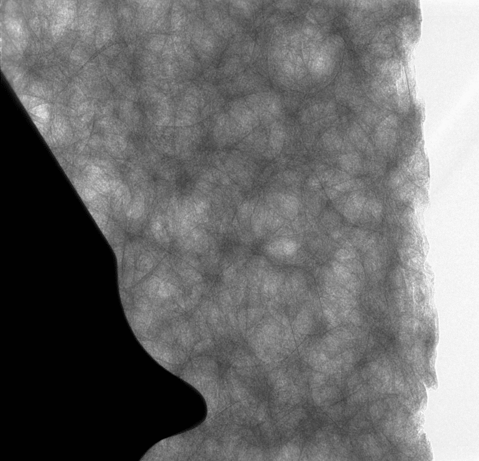
The images show an orthopaedic titanium implant in a bone phantom. Note that the phase contrast improves the visualization of the porous (trabecular) bone structure.
How Detector Technology Shapes Image Quality
Image quality is influenced by every component of an X-ray imaging system, but detector technology plays a fundamental role in determining the information captured during image acquisition. Detector characteristics affect factors such as spatial resolution, sensitivity, field of view, and ultimately the types of samples that can be imaged most effectively.
This philosophy is reflected in the design of the inCiTe™ product family. The original inCiTe™ platform is built around KA Imaging’s BrillianSe™ direct-conversion detector, which supports the system’s phase-contrast imaging capabilities for high-resolution laboratory X-ray microscopy.
With inCiTe™ 2.0, researchers can configure the system with either the BrillianSe™ detector or the Reveal™ detector, allowing the detector to be selected according to the specific imaging requirements of the application. Rather than offering a one-size-fits-all approach, the platform can be tailored to different research workflows while providing access to advanced imaging capabilities.
By treating detector technology as an integral part of the imaging system rather than simply another component, we can design laboratory X-ray microscopy platforms that address a broad range of biomedical research applications.
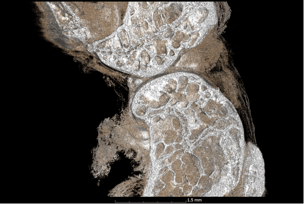
The inCiTe™ 3D X-ray microscope enables high contrast for tissues, like in this mouse stifle joint.
inCiTe™: Phase-Contrast Micro-CT for High-Resolution Imaging
KA Imaging’s inCiTe™ 3D X-ray Microscope applies propagation-based phase-contrast imaging to improve visualization of low-density materials, soft tissues, and the interfaces between different materials. Enhanced edge definition can make structural boundaries more apparent while preserving visualization of mineralized anatomy.
Built around KA Imaging’s BrillianSe™ detector technology, inCiTe™ enables high-resolution laboratory X-ray microscopy designed for research applications involving biomaterials, biological specimens, and other weakly absorbing samples. Detailed 3D imaging can support visualization of internal structures in research specimens without requiring physical sectioning during the imaging workflow.
inCiTe™ 2.0: Combining Phase Contrast and Spectral Imaging
For applications requiring both fine structural detail and improved tissue differentiation, inCiTe™ 2.0 combines phase-contrast and spectral imaging capabilities in a single platform.
This combination helps researchers visualize subtle soft-tissue structures, material interfaces, and compositional differences that may be difficult to distinguish using conventional absorption imaging alone. By combining these complementary contrast methods, inCiTe™ 2.0 combines complementary contrast approaches to provide additional information about complex samples.
Conclusion
Imaging bone and soft tissue together continues to be a fundamental challenge in biomedical research because conventional absorption-based imaging relies on attenuation differences, which inherently provide limited contrast between many soft tissues.
As research questions become increasingly multidisciplinary, imaging systems must do more than produce high-resolution images. They must provide meaningful contrast across complex biological specimens, including the interfaces between hard and soft tissues where many important biological processes occur.
At KA Imaging, our work in detector technology, phase-contrast imaging, and spectral imaging is driven by this objective: helping researchers extract more information from every scan. By combining complementary contrast mechanisms within the inCiTe™ platform, we aim to support biomedical researchers studying the relationships between hard and soft tissues without compromising image quality or workflow efficiency.