
Why Lateral Views Matter
Lateral chest X-rays play an important role in diagnostic imaging. While the frontal (PA) view is the primary assessment, the lateral view adds a second perspective that can help clarify the location, depth, and extent of findings that may be ambiguous on the frontal image alone.
For areas where structures overlap, such as the retrocardiac space behind the heart, the spine, and the posterior costophrenic regions, this alternate view can provide additional anatomical information during image review, supporting clinical decision-making.
What is lateral imaging?
Lateral imaging refers to X-ray views taken from the side of the body, which is different from the standard front-to-back (PA or AP) view.
In chest radiography, lateral views are used alongside frontal views to help localize findings, assess depth, and visualize structures that may be hidden behind the heart, spine, or diaphragm on a frontal image alone, such as the retrocardiac space.
What are the benefits of lateral dual-energy imaging compared to conventional radiography?
Conventional lateral radiographs are limited to a single greyscale image where overlapping bone and soft tissue structures can mask important findings. Lateral dual-energy imaging adds bone and soft-tissue subtracted images to the standard view, helping differentiate overlapping bone and soft-tissue anatomy. This can potentially improve visualization of findings that may be obscured by bone, which shows up opaque on greyscale X-ray images.
Does lateral dual-energy imaging require additional radiation exposure?
No. With Reveal™ 35C, lateral dual-energy images are acquired using the same radiation dose as a conventional chest X-ray, made possible because all three images are captured in a single exposure with no motion artifacts.
When it’s important to pinpoint an exact location—like opportunistically visualizing coronary artery calcifications or confirming where tubes and medical devices are placed—adding a lateral viewpoint can provide additional insight alongside the standard front view.
Why Lateral Views Are Difficult in Traditional Dual-Energy Imaging
Lateral views are difficult in traditional dual-energy imaging because there is a higher chance of motion artifacts and a harder-to-justify dose increase.
Until now, dual-energy has not been commonly used for lateral imaging. Traditional dual-energy technologies involve a two-exposure technique which requires higher radiation and two separate exposures.
This is a particular problem for lateral imaging. Lateral positioning takes longer to set up and is less stable to hold than a standard frontal view, since the patient is rotated 90 degrees and often has less to brace against. With two sequential exposures, even small amounts of patient or cardiac motion between the first and second image can cause misregistration. The bone and soft tissue images don’t line up and appear blurry as a result.
The additional radiation involved is also an issue. Lateral views are often ordered specifically to investigate that retrocardiac region, but doubling the exposure to get a dual-energy lateral image works against the goal of keeping radiation dose proportionate to the diagnostic value of the additional view.
How Reveal 35C Enables Lateral Dual-Energy Imaging
KA Imaging’s Reveal™ 35C, powered by SpectralDR® technology, is designed to address both of the traditional barriers to lateral dual-energy imaging at once.
First, Reveal 35C captures all three images — a standard X-ray, plus supplemental bone and soft tissue dual-energy images — from a single exposure, with zero motion artifacts. Because only one exposure is required,there is no risk of motion misregistration caused by movement between separate exposures. This is a particular advantage for lateral positioning since it’s more difficult to hold a steady position than a standard frontal view.
Moreover, Reveal 35C uses the same X-ray technique, dose, and X-ray source as a standard chest X-ray. This enables lateral dual-energy imaging without requiring additional radiation dose beyond that of a standard chest X-ray exposure.
Because Reveal 35C can be used anywhere a standard X-ray is performed and is compatible with existing fixed, mobile, and portable X-ray systems, this capability can extend to lateral studies in a variety of clinical settings without requiring specialized equipment changes.
Clinical Benefits of Lateral Dual-Energy Imaging
By enabling dual-energy subtraction on lateral views, here’s where Reveal 35C may bring clinical value:
- Improved visualization of overlapping structures: Bone and soft-tissue subtraction on a lateral view can help separate structures that overlap in lateral views.
- Support for opportunistic identification of calcified structures: A lateral dual-energy bone image may improve visualization of coronary and vascular calcifications and support further clinical evaluation.
- Improved visualization of lines, tubes, and devices: Dual-energy subtraction has demonstrated value in improving visualization of lines, tubes, and other overlapping structures that may affect device localization.
- No extra impact on the patient or the workflow: Because Reveal 35C operates within a standard exposure range, lateral dual-energy acquisition is designed to fit into existing imaging protocols without requiring extra exposures or repeat positioning.
Applications for Lateral Dual-Energy Imaging
As an extension of standard lateral radiography, lateral dual-energy imaging may provide additional image information in a variety of clinical scenarios. Potential applications include improved visualization of overlapping anatomical structures and medical devices, select portable imaging workflows, and emerging cardiopulmonary applications where additional anatomical perspective may support image review.
- Pathology differentiation: Lateral dual-energy imaging may provide additional anatomical perspective when overlapping structures make findings difficult to interpret on frontal views alone. By separating bone and soft tissue information while adding a second viewing angle, lateral dual-energy images may help distinguish superimposed anatomical structures from suspicious opacities and support further clinical evaluation.
- Medical device localization: Lateral dual-energy soft tissue images may help clarify the position of lines, tubes, pacemakers, and other hardware such as sternal wires relative to surrounding anatomy.
- Portable and bedside imaging: Because Reveal™ 35C is compatible with fixed, mobile, and portable X-ray systems, lateral dual-energy imaging may also be possible in select portable or bedside scenarios where patient positioning permits.
- Cardiopulmonary assessment: Lateral dual-energy bone images may assist in visualizing coronary artery calcifications as a complement to frontal views.
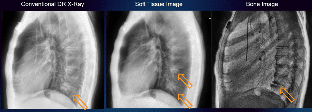
Case example: Differentiating overlapping findings
In one clinical case, a calcified degenerative joint visible on a conventional lateral chest radiograph overlapped with a separate soft-tissue opacity behind the heart. On the conventional image alone, these findings were difficult to distinguish.
The lateral dual-energy soft-tissue image improved separation of soft tissue from overlying bone, making the additional opacity more conspicuous, while the corresponding bone image confirmed the presence of the calcified structure. This case illustrates how lateral dual-energy imaging may provide additional image information when overlapping anatomy complicates interpretation.