
In the intensive care unit (ICU), the chest X-ray comes to the patient. Portable chest X-rays help care teams check lines and tubes and follow changes in the lungs over several days.
Single-exposure dual-energy imaging adds additional information to the chest exam. During one X-ray exposure, the detector records how X-rays of different energies passed through the body. That information can be used to create more than one view of the same anatomy.
How can spectral X-ray imaging fit into an ICU workflow?
When the detector works with the existing portable X-ray system, the exam is positioned and exposed much like a standard bedside chest X-ray. The additional views come from the same exposure and can be reviewed alongside the conventional image.
How are the additional images used in clinical interpretation?
Additional image views may help clinicians see some structures more clearly, but they do not make a diagnosis on their own. Clinicians still interpret the images alongside the patient’s history, exam findings, and other tests.
What is the difference between a conventional X-ray image and dual-energy subtraction images?
A conventional X-ray shows bone and soft tissue overlapping in one grayscale image. Dual-energy images use energy information to reduce the appearance of one type of material. A soft-tissue image reduces the visibility of overlying bone, such as the ribs. A bone image reduces soft tissue and emphasizes bone and calcified structures.
This article looks at how single-exposure spectral imaging can support ICU care, fit into existing bedside workflows, and provide additional information from the same X-ray exposure. Later in the article, we’ll also explore findings from a pilot at Grand River Hospital in Kitchener.
The Traditional ICU Imaging Bottleneck
Bedside chest X-rays are a core part of ICU care. They are fast, they come to the patient, and they allow teams to check on changes as they happen. The challenge is that the ICU sets practical limits on how any image can be taken:
- Some critically ill patients cannot readily be transported to the X-ray room, particularly when they are connected to ventilators, infusion pumps, and monitors. Moving them takes planning, time, and resources.
- Positioning also affects image quality. Portable chest X-rays are usually taken from front to back with the patient lying down or partly sitting up. Patient rotation and smaller lung volumes can make these images harder to interpret than an upright chest X-ray taken in a dedicated room.
- ICU patients often have several lines, tubes, and monitoring leads. These devices overlap with the ribs, the heart, and the lungs. These can become obstructions in certain applications, but X-ray images are also taken to confirm tube and line placement.
- Scatter can reduce image contrast and make some structures harder to distinguish. Anti-scatter grids can improve contrast, but accurate alignment can be challenging during bedside imaging. Their use depends on the equipment and imaging protocol.
When a bedside X-ray does not fully answer the clinical question, the care team may repeat the X-ray, adjust the patient’s position, wait and reassess, or order another test such as CT. Each option has trade-offs. CT may require transporting the patient to a CT suite, depending on the hospital’s configuration.
This raises an important question: Can additional imaging information be captured during the same X-ray exposure already being taken at the bedside?
What Single-Exposure Dual-Energy Imaging Actually Captures
To understand what single-exposure dual-energy imaging adds, let’s look at how X-rays interact with the body.
Why X-ray absorption changes with energy
An X-ray beam contains a range of photon energies. As it passes through the body, some photons are absorbed or scattered, while others reach the detector. This reduction in the X-ray beam is called attenuation.
Attenuation depends on both the material and the energy of the X-rays. At lower energies, materials with higher atomic numbers, such as calcium in bone, absorb X-rays more strongly than soft tissue. The difference is noticeably smaller at higher energies.
A conventional radiograph records the overall attenuation of the X-ray beam in a single image, combining information from the spectrum of X-ray energies. Bone and soft tissue are therefore shown together rather than separated.
From energy information to separate image views
Dual-energy imaging captures information from different X-ray energy ranges. Because materials respond differently at different energies, these measurements can be combined to reduce the visibility of one material relative to another.
This produces complementary image views:
- Standard DR image: Similar to a conventional digital radiograph, showing bone and soft tissue together.
- Soft-tissue image: Reduces the visibility of bone, making soft-tissue structures behind the ribs and clavicles easier to see.
- Bone image: Reduces soft-tissue visibility and emphasizes bone and other dense or calcified structures.
These are not three separate X-rays. They are different image presentations generated from the same imaging event (or exposure). The standard image remains the primary reference, while the additional views provide additional information.
Why single exposure matters
Traditional dual-exposure dual-energy radiography uses two sequential X-ray exposures at different energy levels. Commercial systems using this approach have historically been fixed-room systems, limiting access to dual-energy imaging at the bedside. Because the images are acquired at different times, movement between exposures can also introduce motion or registration artifacts.
Reveal™ 35C takes a different approach. Powered by SpectralDR® technology, its patented multilayer detector captures spectral information from a single standard X-ray exposure, generating conventional, bone-selective, and soft-tissue images.
This single-exposure, detector-based approach brings two important advantages:
- Portability: Spectral information is captured at the detector rather than through a dedicated dual-exposure X-ray system. Reveal 35C can therefore bring dual-energy imaging beyond the fixed radiography room into portable environments, including bedside imaging in the ICU.
- No inter-exposure motion artifacts: Because the spectral information is captured simultaneously, there is no time interval between separate low- and high-energy acquisitions in which the patient can move. This is particularly relevant in settings such as the ICU, where patients may have difficulty remaining still.
By combining single-exposure acquisition with a portable detector, Reveal 35C makes dual-energy X-ray available in clinical environments where traditional dual-exposure systems have not been practical.
How This Fits Into Existing ICU Workflows
Single-exposure spectral imaging is designed to fit into a standard portable X-ray exam. The patient is imaged once, using the existing X-ray system, while the detector captures the information needed to produce both the conventional and spectral images.
What this means at the bedside
Reveal™ 35C is designed to integrate with existing bedside X-ray workflows.
- One exposure: The patient is positioned and imaged once.
- Additional image views: Soft-tissue and bone images are generated from the same exposure.
- Existing equipment: Reveal™ 35C can be used with portable X-ray systems.
- No additional exposures: No additional X-ray exposure is required to generate the spectral image views.
Case Study: Grand River Hospital’s ICU Experience
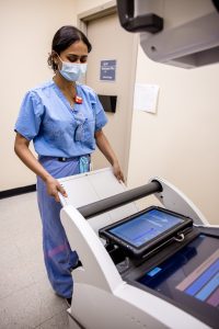
Grand River Hospital in Kitchener, Ontario, partnered with KA Imaging to evaluate Reveal™ 35C in its ICU, including image quality and clinical workflow considerations. The detector was installed on an existing portable X-ray system.
A preliminary analysis compared the six weeks before implementation with the first six weeks of detector use and reported fewer portable chest X-rays and chest CT examinations among ICU patients. The hospital described these as early findings requiring further analysis.
Implementation research presented at the American Society of Emergency Radiology (ASER) Annual Meeting (2024) reported on 104 of 380 ICU chest-X-ray cases using Reveal™ 35C and feedback from 17 clinicians, including nine radiologists and eight intensivists.
Results were positive for more than half of the physicians in all areas, with reported better image quality, no added or faster reading times, increased confidence in diagnosis, and a reduction in time to potential intervention.
Another outcome observed was the potential for CT diversion. In addition to the self-reported reduction trend noted by intensivists, data collected at the hospital showed a decrease in the number of chest CTs during the study period compared to other time frames: a 37.5% decrease compared to the prior 3 months and a 16.67% decline compared to the identical period in the previous year.
Conclusion
Single-exposure spectral imaging adds soft-tissue and bone views to the conventional X-ray without requiring a second exposure. In the ICU, this means additional image information can be available at the bedside during the same imaging event.
The Grand River Hospital experience provides an early example of this approach in clinical practice. Further research can help determine where the additional information provides the most value.