
What is spectral X-ray imaging?
Spectral X-ray imaging uses information from different portions of the X-ray energy spectrum to provide additional information beyond a conventional radiograph.
Reveal™ 35C, powered by SpectralDR® technology, captures spectral information from a single standard X-ray exposure. From that exposure, it generates three images: a conventional DR image, a bone-selective image, and a soft-tissue image.
These complementary images provide clinicians with additional information alongside the conventional radiograph.
You can learn more about how it works here: SpectralDR Technology Explained
How is single-exposure spectral imaging different from traditional dual-energy X-ray approaches?
Dual-energy X-ray uses differences in X-ray energy to differentiate bone and soft tissue. Some traditional dual-energy approaches acquire image information using sequential exposures, which can introduce a time interval between acquisitions.
Reveal™ 35C takes a different approach. Powered by SpectralDR® technology, the detector captures spectral information during a single standard X-ray exposure, enabling dual-energy subtraction without requiring a second exposure.
The result is a conventional DR image plus supplemental bone and soft-tissue images acquired simultaneously.
Does spectral imaging require a higher radiation dose?
With Reveal™ 35C, no additional exposure is required to generate the spectral images. The conventional, bone, and soft-tissue images are all generated from a single standard X-ray exposure.
Reveal™ 35C is designed to provide single-exposure spectral X-ray without added dose or workflow changes compared with standard DR imaging.
What additional information can spectral images provide compared with a conventional X-ray?
A conventional X-ray combines information from bone, soft tissue, and other structures into a single image. Overlapping anatomy can make some findings more difficult to visualize.
The supplemental bone and soft-tissue images generated by Reveal™ 35C provide complementary information that can help improve visualization of clinically relevant features that may be partially obscured by overlapping anatomy on the conventional image.
Where can spectral X-ray imaging be used?
Reveal™ 35C is a portable spectral X-ray detector designed to support imaging across clinical environments. It can be used as a retrofit detector with compatible X-ray systems and can also be integrated into mobile and portable imaging systems.
This makes single-exposure dual-energy imaging available across settings including the ICU, emergency room, cancer center, cardiac care, long-term care, and urgent care.
The Challenges of Single-Image, Single-Energy X-ray
Conventional single-image, single-energy X-ray is widely used across healthcare because of its speed, accessibility, and established role in clinical imaging.
A conventional radiograph combines information from bone, soft tissue, and other structures into a single grayscale image. Because these structures overlap, some clinically relevant features can be more difficult to visualize.
For example:
- A pulmonary finding overlapped by a rib may be more difficult to visualize on a conventional image.
- A line or tube tip may be more difficult to visualize when it overlaps surrounding anatomy.
- Calcifications, retained surgical objects, or subtle soft tissue findings may be less conspicuous when superimposed with surrounding anatomy.
Dual-energy X-ray provides another way to look at this overlapping information.
How Dual-Energy Addresses This Challenge
Dual-energy X-ray takes advantage of differences in how bone and soft tissue attenuate X-rays at different energies. This information can be used to generate separate bone and soft-tissue images alongside a conventional radiograph.
Historically, some dual-energy approaches have relied on sequential acquisitions. Because the images are acquired at different points in time, patient motion between exposures can result in motion or alignment artifacts.
Single-exposure dual-energy imaging provides a different approach by acquiring the information needed for bone and soft-tissue separation simultaneously.
Reveal 35C: How Our X-ray Detector Captures Three Images From One Exposure
KA Imaging’s Reveal™ 35C is a portable spectral X-ray detector powered by SpectralDR® technology. SpectralDR® enables dual-energy subtraction from a single standard X-ray exposure, generating three images simultaneously: a conventional DR image, a bone image, and a soft-tissue image.
Reveal™ 35C uses a patented triple-layer detector design to capture spectral information at the detector level during the X-ray exposure. The resulting information is used to differentiate bone and soft tissue.
Because the image information is captured from the same exposure, Reveal™ 35C generates the conventional, bone, and soft-tissue images without the motion artifacts associated with sequential dual-exposure acquisition.
Reveal™ 35C is available as a retrofit detector for compatible X-ray systems and can also be integrated into mobile and portable imaging systems.
Bone, Soft Tissue, and Conventional: What Each Spectral Image Shows
A single exposure with Reveal™ 35C produces three complementary image types:
| Image Type | What It Shows | May Help With |
|---|---|---|
| Conventional (DR) image | The standard grayscale composite combining bone and soft-tissue information | Provides an anatomical overview with bone and soft-tissue structures shown together |
| Soft tissue image | Reduces bone visibility, with emphasis on soft tissue structures | Supporting visualization of masses, nodules, or opacities that may be partially obscured by overlapping anatomy |
| Bone image | Removes soft tissue, with emphasis on skeletal and calcified structures | Supporting visualization of calcifications, line and tube tips, and retained surgical or medical devices |
Rather than replacing the conventional radiograph, the bone and soft-tissue images provide complementary information alongside it.
For example, when a finding overlaps a rib or other bony anatomy on the conventional image, the soft-tissue image may provide a clearer view of that region. The corresponding bone image provides complementary information about skeletal and calcified structures captured during the same exposure.
The following clinical example demonstrates how these three images can provide different views of the same anatomy.
Clinical Example: Conventional DR and Spectral Images Acquired with Reveal™ 35C.
All three images in this example were generated from a single X-ray exposure with Reveal™ 35C.
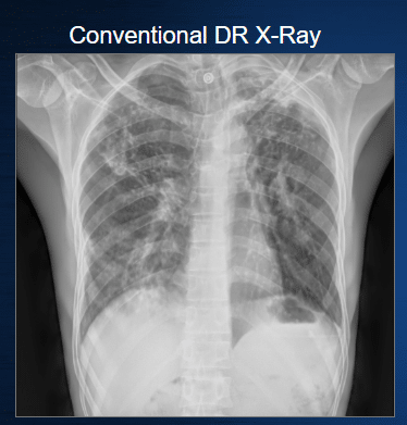
Conventional DR combines bone and soft tissue structures into a single image. In this clinical example, overlapping anatomy illustrates how additional spectral information can provide a complementary view of the lung.
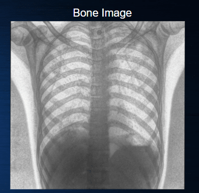
The bone image emphasizes skeletal and calcified structures while reducing the visual contribution of surrounding soft tissue, providing a complementary view of the anatomy captured in the same exposure.
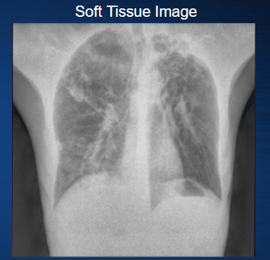
The soft tissue image reduces the visual impact of overlapping bone, providing a complementary view of the lung anatomy. The lung apices are well visualized, where overlapping ribs and other structures can otherwise obscure underlying pathology.
Note, again, that all three images were produced from a single exposure.
In this case, the patient was subsequently referred for GeneXpert testing to confirm tuberculosis. The example illustrates the complementary visualization provided by the conventional, bone, and soft-tissue images.
Why This Doesn’t Require Extra Exposures
Generating three images with Reveal™ 35C does not require three X-ray exposures.
SpectralDR® technology captures the spectral information needed to generate the conventional, bone, and soft-tissue images from a single standard X-ray exposure. Reveal™ 35C therefore provides supplemental dual-energy images without added dose or workflow changes compared with standard DR imaging.
Single-exposure acquisition also means there is no time interval between separate acquisitions for patient movement to create the motion artifacts associated with sequential dual-exposure imaging.
The result is straightforward: one exposure, three complementary images.
Reveal™ 35C, powered by SpectralDR® technology, brings single-exposure spectral X-ray imaging to portable, retrofit, and fixed imaging environments—providing conventional, bone, and soft-tissue images without requiring a change to the standard X-ray workflow.