
Since X-rays first began examining soft tissue in the early 20th century, CXR has played a pivotal role in diagnosing pneumonia. Early radiographs were fairly rudimentary compared to modern standards, but they allowed physicians to see pneumonia-associated changes in lung fields. In the last 100 years since its discovery, X-ray lacked any major technological advancements until the introduction of SpectralDR, developed by KA Imaging. Our team at KA Imaging has remained trailblazers in the radiography landscape, innovating imaging technology to new heights. This article explores the importance of enhanced CXR for pneumonia, and how KA Imaging’s patented Reveal 35C X-ray Detector with SpectralDR technology innovates the standards for accurate CXR.
The Importance Of Accurate CXR For Pneumonia
Pneumonia often presents with infiltrates, which are areas of increased density (opacity) in the lung tissue due to infection and inflammation. What makes enhanced CXR for pneumonia so important is that it helps not just diagnose, but also determine the extent and location of the pneumonia. This includes identifying whether it is affecting a single lobe (lobar pneumonia), multiple lobes (multilobar pneumonia), or diffusely spread across the lungs (bronchopneumonia). This increased level of knowledge in the pneumonia’s severity has a major impact in how medical personnel administer life-saving treatment. Moreover, radiographic visualization is sometimes able to detect complications caused by pneumonia, such as empyema or abscess formation.
Based on the CXR findings, medical personnel can decide whether further testing is necessary. Our research has found that there are many cases in which our X-ray technology alone can identify pneumonia in patients. Diagnosis solely using a single X-ray examination when possible is beneficial for both patients and medical facilities.
Symptoms of pneumonia can overlap with other chest conditions such as congestive heart failure, pulmonary edema, lung abscesses, or malignancies. This is where intensive CXR visualization and accuracy becomes most important. Accurate CXR helps differentiate pneumonia from these other possibilities through visualization of unique, characteristic lung patterns.
Reveal with SpectralDR technology: The World’s First Portable Dual-Energy Detector
As CXR plays such an important role in pneumonia diagnosis, radiographic technology developers have worked on improving aspects such as image quality or technology deployment (like with mobile systems). But essentially, digital radiography has remained fairly the same. With KA now introducing revolutionary imaging technology that goes beyond just image quality and resolution, various conditions that are visualized in radiography are easier to visualize, examine, and therefore, diagnose. Quick diagnosis unlocks the potential for even faster medical treatment.
KA Imaging’s mission is to empower limitless innovation for radiographic imaging, simplifying and supporting medical testing efforts to improve detailed identification of life-threatening conditions, including pneumonia.
The Reveal 35C X-ray Detector
KA Imaging developed the world’s first portable single exposure dual-energy detector, the Reveal 35C X-ray Detector. This patented technology is called SpectralDR. Reveal 35C is capable of improving the visualization of many conditions through radiographic imaging, but is most known for its good results achieved in detecting signs of pneumonia with CXR. Let’s dive into how Reveal 35C’s technology enhances radiographic visualization.
Dual-Energy Subtraction (DES): The Old Ways vs. SpectralDR technology
Old approaches of DES technology involve taking two separate X-ray exposures at different energy levels (usually a high-energy and a low-energy exposure). The difference in absorption of X-rays by various tissues at these two energy levels provides a basis for separating images of different tissue types.
KA Imaging developed a patented cutting edge DES technology known as SpectralDR. SpectralDR is unique from other DES technology because it acquires multiple energies in one CXR scan while not requiring increased radiation doses. The standard radiation dose needed for the average CXR is all that is needed for Reveal’s technology to produce crisp imaging. Additionally, this imaging can be executed with no distortion or errors caused by motion.
Triple Stacked Layer Design
Single exposure dual-energy subtraction X-ray – our SpectralDR technology – is only possible because of Reveal’s distinctive triple stack design. Thanks to SpectralDR, our mobile X-ray detector is capable of simultaneously acquiring three images with only one exposure. The technology is capable of producing separated imaging on bone or soft tissue, improving clarity and reducing the chances of important visualization being obstructed.
How Reveal Technology Highlights Visible Signs Of Pneumonia
Thanks to its innovative 3-in-1 design, Reveal 35C is capable of capturing diverse imaging of the subject with just a single exposure. It can capture images of bones and tissues, together and individually. This means that doctors will obtain imaging that focuses both on the soft tissue, and alternatively the bone surrounding it, in the case of a CXR for pneumonia, the ribs and lungs. With Reveal 35C, doctors can effectively capture and visually differentiate lung nodules, ribs fractures, and even foreign objects within the body with ease.
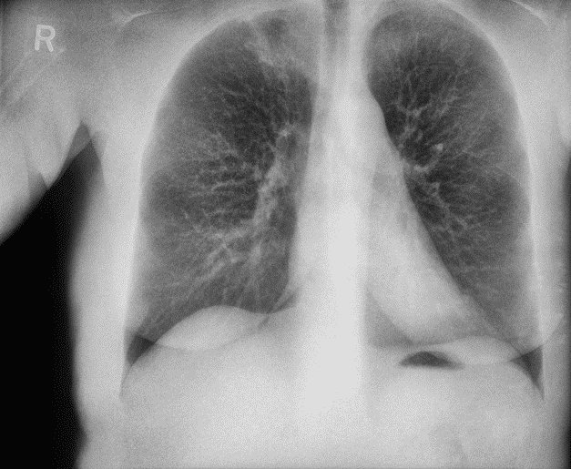
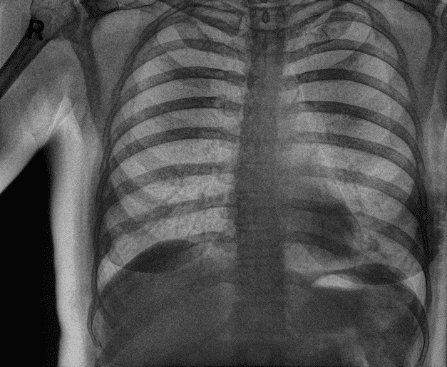
On the left, the soft tissue is visible with no rib imaging to obstruct it. In the middle image, the Reveal 35C produces distinct exposure of the rib bones.
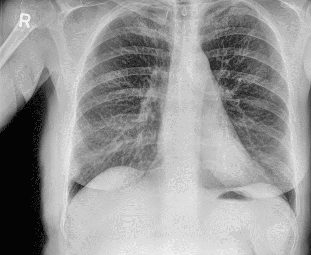
Reveal 35C’s 3-in-1 technology provides three distinct photos of the subject, a standard image of the ribs and lungs, a photo highlighting just the ribs, and a photo visualizing beyond the ribs so that only the lungs are visible.
In a paper presented at RSNA in 2021, the Reveal 35C technology was shown to detect 33% more pneumonia cases (including COVID-19) than traditional X-Ray. The paper presents the initial results of an ongoing clinical trial that is being carried out in Toronto (Canada).
With Reveal 35C, medical personnel can overcome visual obstructions caused by bone, in this case the ribs, to get a better view of the soft tissue, the lungs. This makes it possible to examine signs of pneumonia that would otherwise be obstructed and unviewable from behind the ribs. This has allowed for quicker identification and improved accuracy in diagnosis when performing CXR for pneumonia. As previously mentioned, this not only applies to diagnosis, but also examination of the pneumonia’s severity and appearance of side effects. The quicker the pneumonia and its side effects are identified and diagnosed, the quicker medical personnel are able to administer adequate treatment. If a single x-ray can identify pneumonia and its side effects, medical personnel can potentially avoid the need for further imaging, speeding up treatments and saving on cost, time, and resources.
Reveal 35C is the first single exposure dual-energy X-ray detector to deliver a portable imaging solution, only sold by KA Imaging. Mobile imaging technology is becoming a necessity for medical personnel, as it allows more flexibility in emergency situations, long-term care homes, or when examining patients who cannot move easily like in the ICU. Medical facilities aim to limit the transportation of highly infectious patients in order to avoid contaminating additional spaces within the facility. Mobile X-ray imaging allows for doctors to assess patients from their bedside, reducing the risk of constant movement of patients with contagious conditions, such as COVID-19. Reveal 35C is the first X-ray detector capable of capturing single exposure dynamic imaging in mobile use cases, expanding the capabilities of doctors and the comfort they can provide for their patients.
At KA Imaging, our team is focused on innovating imaging technology with innovation of our phase contrast and Spectral DR features. Learn more about Reveal 35C’s capabilities, how to purchase a Reveal 35C for your medical facility, and what’s next for KA Imaging’s X-ray technology.
The importance of accurate CXR in identifying the location, extent, and complications of pneumonia cannot be overstated. KA Imaging’s Reveal 35C, with its patented Dual-Energy Subtraction (DES) and triple-stacked layer design, marks a revolutionary advancement in CXR for pneumonia. By providing clear, obstruction-free images with a single exposure, Reveal 35C aids precise and speedy diagnosis, critical for effective patient care. Its portability further enhances its utility, especially in point-of-care settings where rapid, accurate diagnostics are paramount. Investing in innovative solutions like the Reveal 35C not only improves diagnostic accuracy for pneumonia but also elevates the overall standards of medical care, ultimately saving lives and enhancing patient treatment.