Entries by Fernanda Fraga

, ,

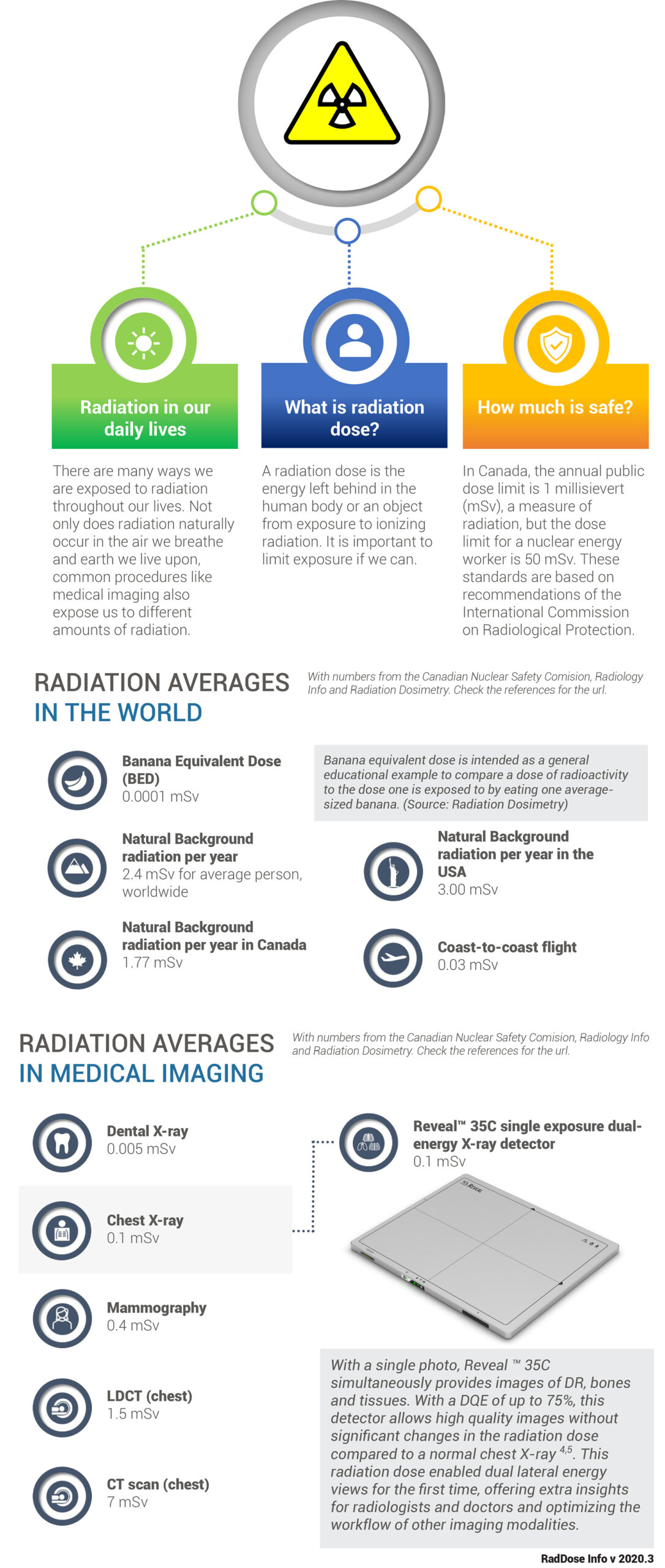
X-ray After 125 Years

The Discovery

illustration of Wilhelm RöntgenIn 1895, German physicist Wilhelm Röntgen was studying electron beams when he made an accidental discovery4. He noticed that when he covered a Crookes tube (an early vacuum tube that produces electron beams) with cardboard, a faint glow came from a screen painted with fluorescent material that was some distance away, further than the electron beam could travel. Next, he realized that objects placed between the tube and the screen appeared transparent, although some more than others, depending on the material. He deduced that invisible rays other than the electron beam were coming from the tube and named them X-rays, due to their unknown nature. He was able to demonstrate his discovery to the world through the world’s first X-ray image that he took of his wife’s hand on a photographic plate that showed the bone structure within.

After significant advancements, at a time where people were becoming more excited about advancing technology, X-rays became a huge fad. Machines were used in shoe stores to check sizing from the 1920s to 1960s, and a hand-held toy was even made so children could take their own images at home. The dangers of radiation soon became evident after the use of nuclear bombs in World War II and exposure to X-rays was restricted to certain controlled applications.

The Basics

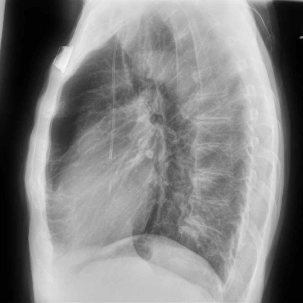
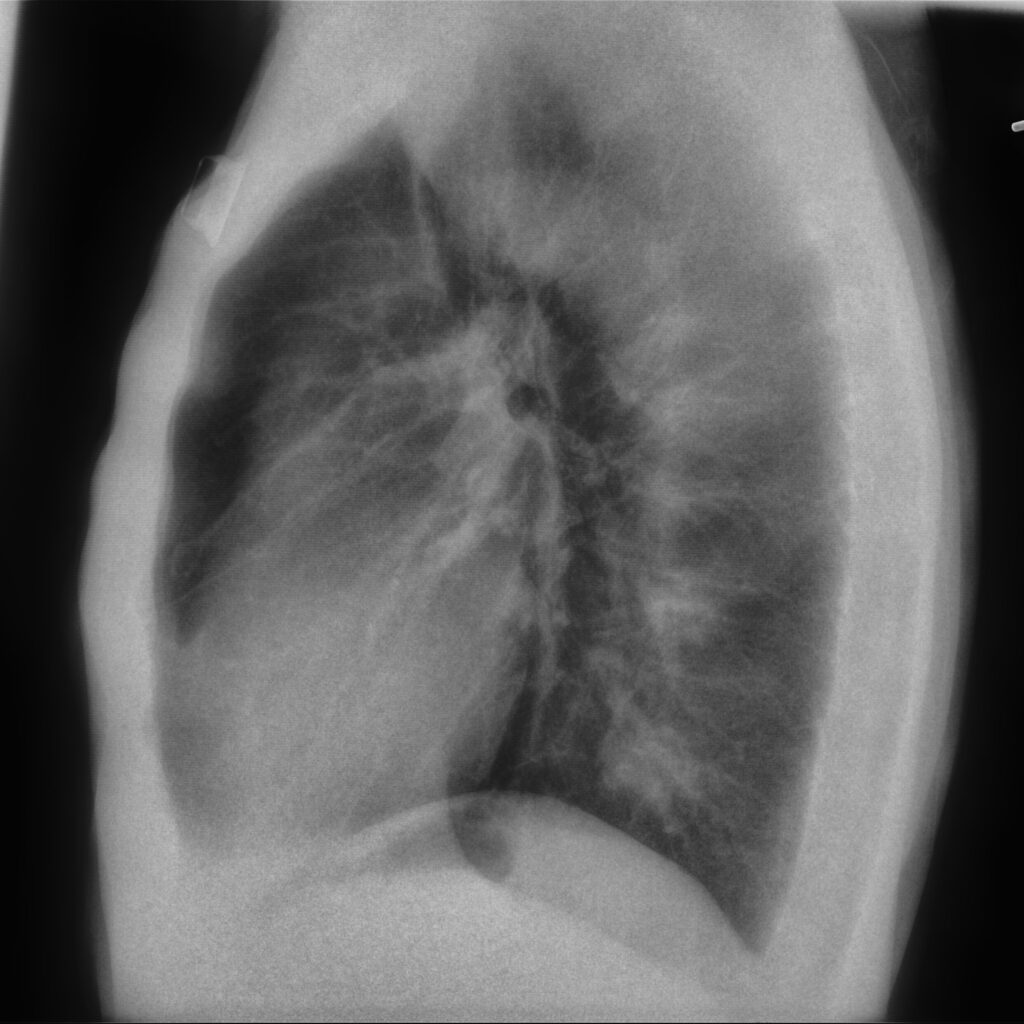
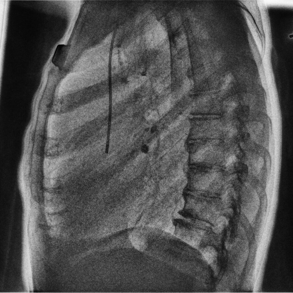
We understand now that X-rays are a type of radiation that can pass through the human body, and the rays are invisible and intangible. As they pass through an object, their energy is absorbed at different rates by different components within the object due to different atomic numbers. A detector on the other side of an object or body can transform these X-rays into an image to view and process.

Objects within a human body such as bones require higher energy X-rays to penetrate, which is why they appear clearly in the images. Softer tissue appears darker, as X-rays travel more easily through less dense materials with low atomic numbers like fat or muscle and air-filled cavities such as the lungs.

Applications

The main application of X-ray imaging has continued to be in the medical field, though X-ray imaging is also used for industrial, scientific and veterinary purposes. Röntgen, along with his colleagues, realized the importance of his discovery immediately and proceeded to study and experiment with enthusiasm.

We know a great deal more about X-rays now due to their persistence and have been able to integrate the use of X-rays into diagnostic procedures and medical care.

X-ray imaging is a cost-effective primary triaging and diagnostic tool for medical practitioners and helps to provide efficient care to patients presenting with illness or injury. It is a valued tool for a wide variety of examinations like diagnosing lung diseases or monitoring recovery, detecting bone fractures, supporting medical treatments, and guiding personnel as they insert devices into a patient’s body, or even treatment planning for cancer treatment.

Risks and Benefits

Of course, there have always been some risks, but these are much less threatening in modern times. X-ray imaging uses ionizing radiation, which potentially has enough energy to cause damage to DNA. There is a small chance that a person may develop cancer later in life, according to the FDA2, or have tissue damage causing cataracts, skin reddening or hair loss. These side-effects are extremely rare and depend on other variables and amount of radiation exposure.

The potential benefits of using X-ray imaging provide balance to these risks, and steps are taken to maintain the safest environment for patients and practitioners. Special care should be provided for children, pregnant women and those who are known to be more sensitive to radiation.  


Moving forward into the future

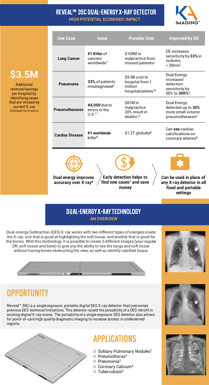
Areas of focus moving forward continue to be reducing radiation dose, improving image resolution, and enhancing materials and methods. Emerging companies like KA Imaging have taken strides in these areas and are producing new technology to support medical image quality and design by leveraging all parts of the X-ray spectrum. Reveal™ 35C, for example, brings new opportunities for portable, high-quality imaging without motion artifacts, capable of distinguishing between bone and soft tissue in one single shot, and improvements in X-ray dose by lowered exposure to the patient.


References

  1. Britannica – https://www.britannica.com/science/X-ray/Fundamental-characteristics

Reveal 35C Featured in FIND Report on Tuberculosis Screening

KA Imaging Inc. was featured in a report about digital chest radiography and computer-aided detection solution by Foundation for Innovative New Diagnostics. The Foundation for Innovative New Diagnostics (FIND) explores the market of radiology equipment and solutions for screening TB1, a disease that is ranked top 10 cause of deaths worldwide2

The purpose of the report is to help readers understand the different technologies used in performing chest X-ray radiography along with the progress of diagnostic X-ray imaging. Reveal™ 35C X-ray detector is the world’s first dual-energy detector, enabling bone and soft-tissue differentiation in one single exposure without motion artifacts. This new technology allows for better visualization, is highly affordable and can easily upgrade fixed and portable settings.  

Foundation for Innovative New Diagnostics partnered with World Health Organization and Stop TB to raise more awareness of the Tuberculosis epidemic. Nearly 4,000 people dies from TB every day1 but is unrecognized as the COVID-19 pandemic remains in the headline a year after.  

REFERENCES

1. Digital Chest Radiography and Computer-Aided Detection (CAD) Solutions for Tuberculosis Diagnostics. Foundation for Innovative New Diagnostics. Retrieved March 30, 2021 from https://www.finddx.org/wp-content/uploads/2021/03/Digital-CXR-CAD-solutions-for-TB-diagnosis-24Mar2021.pdf 

2. World Health Organization. (2020). Global Tuberculosis Report. Retrieved March 18, 2021 from https://apps.who.int/iris/bitstream/handle/10665/336069/9789240013131-eng.pdf 

March 24: World Tuberculosis Day

March 24 is World Tuberculosis Day. The date is celebrated to raise public awareness of all TB impacts. This disease has serious health, social and economic consequences. Find out more in the infographic below:

  1. World Health Organization. (2020). Global Tuberculosis Report. Retrieved 18, 2021 from https://apps.who.int/iris/bitstream/handle/10665/336069/9789240013131-eng.pdf
  2. World Health Organization. (2020). The End TB Strategy. Retrieved March 19, 2021 from https://www.who.int/tb/strategy/en/
  3. World Health Organization. (2015). Global strategy and targets for tuberculosis prevention, care and control after 2015. Retrieved March 18, 2021 from https://www.who.int/tb/post2015_TBstrategy.pdf?ua=1

Upgrade To Reveal 35C: an economic Proposition

Click here to fill the form and receive the PDF.


Fill the form to unlock the PDF.


KA Imaging is committed to protecting and respecting your privacy, and we’ll only use your personal information to administer your account and to provide the products and services you requested from us. From time to time, we would like to contact you about our products and services, as well as other content that may be of interest to you. If you consent to us contacting you for this purpose, please tick below to say how you would like us to contact you:


You can unsubscribe from these communications at any time. For more information on how to unsubscribe, our privacy practices, and how we are committed to protecting and respecting your privacy, please review our Privacy Policy.

By clicking submit below, you consent to allow KA Imaging to store and process the personal information submitted above to provide you the content requested.

UPGRADE TO REVEAL 35C: AN ECONOMIC PROPOSITION


Dual energy has been shown to detect more solitary pulmonary nodules, pneumothorax, pneumonia, tuberculosis and coronary calcium than conventional analog or digital chest X-ray. This early detection enables better patient outcomes, additional hospital revenue, cost savings to healthcare systems and reduces exposure to malpractice claims.

KA Imaging has created Reveal™ 35C, the world’s first digital portable dual-energy X-ray detector as a universal upgrade for any analog or digital X-ray detector from any manufacturer. 

The infographic below summarizes the study.

UPGRADE TO REVEAL 35c: AN ECONOMIC PROPOSITION

Fill the form to receive the complete study in your email.


References

1.Oda, Seitaro, Kazuo Awai, Yoshinori Funama, Daisuke Utsunomiya, Yumi Yanaga, Koichi Kawanaka, Takeshi Nakaura et al. “Detection of small pulmonary nodules on chest radiographs: efficacy of dual-energy subtraction technique using flat-panel detector chest radiography.” Clinical radiology 65, no. 8 (2010): 609-615.

2.Martini, Katharina, Marco Baessler, Stephan Baumueller, and Thomas Frauenfelder. “Diagnostic accuracy and added value of dual-energy subtraction radiography compared to standard conventional radiography using computed tomography as standard of reference.” PloS one 12, no. 3 (2017): e0174285.

3.Urbaneja, A., Dodin, G., Hoosu, G., et al. (2018) Added Value of Bone Subtraction in Dual-energy Digital Radiography in the Detection of Pneuomothorax: Impact of Reader Expertise and Medical Specialty. The Association of University Radiologists. Elsevier Inc.

4. Song, Yingnan, Hao Wu, Di Wen, Bo Zhu, Philipp Graner, Leslie Ciancibello, Haran Rajeswaran et al. “Detection of coronary calcifications with dual energy chest X-rays: clinical evaluation.” The International Journal of Cardiovascular Imaging (2020): 1-8.

5.Sharma, Madhurima, Manavjit Singh Sandhu, Ujjwal Gorsi, Dheeraj Gupta, and Niranjan Khandelwal. “Role of digital tomosynthesis and dual energy subtraction digital radiography in detection of parenchymal lesions in active pulmonary tuberculosis.” European Journal of Radiology 84, no. 9 (2015): 1820-1827.

6.Kuhlman, Janet E., Jannette Collins, Gregory N. Brooks, Donald R. Yandow, and Lynn S. Broderick. “Dual-energy subtraction chest radiography: what to look for beyond calcified nodules.” Radiographics 26, no. 1 (2006): 79-92.

7.https://www.who.int/news-room/fact-sheets/detail/cancer

8.Song, Yingnan, Hao Wu, Di Wen, Bo Zhu, Philipp Graner, Leslie Ciancibello, Haran Rajeswaran et al. “Detection of coronary calcifications with dual energy chest X-rays: clinical evaluation.” The International Journal of Cardiovascular Imaging (2020): 1-8.

9.https://www.who.int/health-topics/cardiovascular-diseases#tab=tab_1

10.Divino, V., etal “The annual economic burden among patients hospitalized for community-acquired pneumonia (CAP): a retrospective US cohort study.” Current Medical Research and Opinion 36, no. 1 (2020): 151-160.

11.https://www.healthgrades.com/quality/patient-safety-2020-infographic

12.Domino etal, “Injuries and Liability Related to Central Vascular Catheters–A Closed Claims Analysis”, Anesthesiology 2004; 100:1411-8.

* Contact us to see the full study

,

KA Imaging to Present at the 31st Annual Oppenheimer Healthcare Conference

WATERLOO, ON (March 15, 2021) – KA Imaging, a company that develops innovative X-ray imaging solutions, announced that management will deliver a company presentation at the 31st Annual Oppenheimer Healthcare Conference on Tuesday, March 16, 2021 at 8:00 a.m. ET.

The virtual presentation will be broadcast live and archived on the Company’s website at under “News and Events” in the About Us section.

About KA Imaging

A spin-off from the University of Waterloo, KA Imaging specializes in developing innovative X-ray imaging technologies and systems, providing solutions to the medical, veterinary, and non-destructive test industrial markets. For more information, please visit www.kaimaging.com.

Contact:

Stephen Kilmer

Investor Relations

skilmer@kaimaging.com

T: 647.872.4849

The Detection of Pneumothorax and How Dual-Energy Can Help

Click here to fill the form and receive the PDF.


Fill the form to unlock the PDF.


KA Imaging is committed to protecting and respecting your privacy, and we’ll only use your personal information to administer your account and to provide the products and services you requested from us. From time to time, we would like to contact you about our products and services, as well as other content that may be of interest to you. If you consent to us contacting you for this purpose, please tick below to say how you would like us to contact you:


You can unsubscribe from these communications at any time. For more information on how to unsubscribe, our privacy practices, and how we are committed to protecting and respecting your privacy, please review our Privacy Policy.

By clicking submit below, you consent to allow KA Imaging to store and process the personal information submitted above to provide you the content requested.

how dual-energy x-ray can help detect pneumothorax


Dual-energy bone subtraction radiographs yielded the greatest increase in detection, even when used alone. The study reported the positive result in diagnostic performance with a sensitivity and specificity for pneumothorax detection as 82% and 92%, compared to 70% and 84% in sensitivity and specificity for conventional radiographs. The physicians in this case recommended the use of dual-energy bone subtraction for suspected pneumothorax without hesitation.

Read the blog post below for a summary of the article.


Pneumothorax, or a collapsed lung that occurs when air enters the space around the lungs, is a condition that poses diagnostic problems. Although common tests, such as lung sonography, ultrasound, CT scan and X-ray are often performed, some cases may be misdiagnosed. This could present a life-threatening situation due to the immediate treatments often needed for this medical emergency.

Several studies on patient safety look at iatrogenic (caused by medical procedure) pneumothorax in American hospitals. Healthgrades 2020 Patient Safety Excellence Award infographic reported that from 2016-2018 there were more than 44,000 Medicare patients in U.S. hospitals diagnosed with a collapsed lung due to a procedure in or around the chest alone. There are also at least 10,000 cases per year in Canada2.

A recent study in France, published in 2018, calls the condition common and reports 17 out of 100,000 people are affected there, with 34.8 out of 100,000 people in England estimated to be affected by pneumothorax as well4.

A team of doctors studied the detection of pneumothorax and the value of using dual-energy digital radiography, as well as the impact of medical specialty. They looked at forty patients with a suspected pneumothorax and divided them into groups based on their final diagnosis – whether they were thought to have this issue or not. They were able to compare and analyze standard images, dual-energy bone subtracted images and a combination in specific sessions. Doctors with various levels of expertise were included in the study.

They found that bone subtracted dual-energy thoracic radiographs improved the detection of pneumothorax, including small pneumothoraxes, regardless of the medical specialty or reader’s level of expertise.

In fact, dual-energy bone subtraction radiographs yielded the greatest increase in detection, even when used alone. The study reported the positive result in diagnostic performance with a sensitivity and specificity for pneumothorax detection as 82% and 92%, compared to 70% and 84% in sensitivity and specificity for conventional radiographs4. The physicians in this case recommended the use of dual-energy bone subtraction for suspected pneumothorax without hesitation.

Dual-energy radiography is an expanding technique and has demonstrated improvement in the detection of calcified and noncalcified lung nodules, and calcified thoracic lesions, mediastinal, pleural, or pulmonary. This technique supresses bone structures of the thoracic wall, reduces superimposition and allows for finer delimitation of lung contours.

KA Imaging’s Reveal, a dual-energy X-ray detector that has received USA FDA 510 (k) clearance as well as a Health Canada Medical Device Licence, is a unique and fitting solution to help in the diagnostic process. 

Reveal is a portable detector that enables bone and soft-tissue differentiation without motion artifacts in a single X-ray exposure. It can replace existing X-ray detectors and is also retrofittable to existing X-ray systems. Reveal increases sensitivity compared to conventional X-ray to aid in the early diagnosis and monitoring of pulmonary conditions. It is much more affordable than other fixed dual-energy systems, at less than a quarter of the price, and can be taken right to a patient’s bedside in an emergency room setting.

Reveal uses significantly less radiation than a CT and provides clearer images than a conventional X-ray. In one shot, the detector simultaneously delivers DR, bone, and tissue images.

References

  1. Right Diagnosis from Healthgrades. Statistics about Pneumothorax. https://www.rightdiagnosis.com/p/pneumothorax/stats.htm
  2. Healthgrades. Patient Safety 2020 Infographic. https://www.healthgrades.com/quality/patient-safety-2020-infographic
  3. Reed, K. May, R., et al. (2011) Healthgrades Patient Safety in American Hospitals Study. HealthGrades, Guiding America to Better Healthcare. https://patientsafetymovement.org/wp-content/uploads/2016/02/Resources_Reports_Patient_Safety_in_American_Hospitals_Study.pdf
  4. Urbaneja, A., Dodin, G., Hoosu, G., et al. (2018) Added Value of Bone Subtraction in Dual-energy Digital Radiography in the Detection of Pneuomothorax: Impact of Reader Expertise and Medical Specialty. The Association of University Radiologists. Elsevier Inc.

,

KA Imaging Further Strengthens Executive Team with Appointment of World-Renowned Radiologist, Dr. Philip Templeton, as Chief Medical Officer

A board certified radiologist, Dr. Templeton brings over 38 years of academic and entrepreneurial medicine experience to KA Imaging. Dr. Templeton is most recently known as Cofounder and Chief Medical Officer of DocPanel Technologies. A graduate and valedictorian at the University of Rochester School of Medicine, he did his internship in Medicine at the Georgetown University Hospital. His radiology residency was at the University of Maryland, where he also served as Chief Resident. He undertook his Thoracic Radiology Fellowship at the Massachusetts General Hospital. He served as Professor and Chairman of University of Maryland Radiology for ten years. Dr. Templeton was inducted as a Fellow of the American College of Radiology in 2000.

Among his many career accomplishments, Dr. Templeton was a pioneer in the use of several groundbreaking technologies, including CT fluoroscopy, digital X-ray, PACS and teleradiology.

“In addition to being a skilled physician, Dr. Templeton is a seasoned corporate leader with a strong track record of delivering medical, scientific and strategic guidance to both established and emerging companies in the medical imaging space,” said Amol Karnick, CEO of KA Imaging. “We are delighted to welcome him as CMO. Dr. Templeton will provide a strong liaison with our customers, and ensure that our clinical programs are designed to meet the needs of both clinicians and patients.”

Dr. Templeton’s appointment comes on the heels of the recent additions of two other seasoned industry veterans – Shawn Campbell as Vice President of Operations and Robert Moccia as Vice President of Sales, USA and Canada – as KA Imaging executes its commercial roll-out of Reveal™ 35C, the world’s first portable, single exposure, dual-energy X-ray detector that enables bone and soft tissue differentiation without motion artifacts.

About KA Imaging

A spin-off from the University of Waterloo, KA Imaging specializes in developing innovative X-ray imaging technologies and systems, providing solutions to the medical, veterinary, and non-destructive test industrial markets. For more information, please visit www.kaimaging.com.

Contacts

Fernanda Fraga
Media Relations
ffraga@kaimaging.com
T: 226.215.9897

Stephen Kilmer
Investor Relations
skilmer@kaimaging.com
T: 647.872.4849

,

KA Imaging to Present Poster on unprecedented Lateral Dual-Energy X-ray Views at ECR 2021

WATERLOO, ON (February 24, 2021)Business Wire – KA Imaging, a company that develops innovative X-ray imaging solutions, announced that a poster reporting on results from a study examining the feasibility of lateral chest dual-energy subtraction radiography using a stacked single-exposure multi-layer x-ray detector will be presented by Sebastian Maurino, a medical physicist at KA Imaging, at this year’s European Congress of Radiology (ECR).As highlighted in the poster, KA Imaging’s novel approach allows radiologists to take full advantage of lateral dual-energy images by obtaining both conventional and dual–energy subtraction lateral images simultaneously, thereby making it considerably easier to differentiate between materials behind the heart.

Quoting directly from the poster, “The DE lateral image also visualizes calcified joints and masses behind the heart that are not visible in the [Posterior-Anterior (PA)] image. Multi-layer detectors were shown to provide DE PA and lateral images of the chest at conventional patient dose levels.”

“Older generation dual-energy subtraction X-ray machines need two X-ray exposures. This results in considerable additional radiation exposure for the patient, helping to explain why lateral dual-energy images are so uncommon today,” said Karim S Karim, Ph.D., CTO of KA Imaging. “KA Imaging’s proprietary technologies have eliminated these extra radiation concerns by working with only one single standard chest X-ray exposure that has the added benefit of zero motion artifacts.”

ECR, being held virtually this year from March 3 – 7, is one of the largest medical meetings in Europe and the second-largest radiological meeting in the world. ECR attendees span all areas of the radiology arena including: radiology professionals, radiographers, physicists, industry representatives, and press reporters for both the medical and consumer press. In addition to participating in the scientific program of ECR 2021, KA Imaging will also host a virtual exhibition booth throughout the meeting.

DR

Soft tissue image

Bone image

About KA Imaging

A spin-off from the University of Waterloo, KA Imaging specializes in developing innovative X-ray imaging technologies and systems, providing solutions to the medical, veterinary, and non-destructive test industrial markets. For more information, please visit www.kaimaging.com.

Contacts:

Fernanda Fraga 
Media Relations
ffraga@kaimaging.com 
T: 226.215.9897

Stephen Kilmer
Investor Relations
stephen@kilmerlucas.com
T: 647.872.4849

Detecting Small Pulmonary Nodules

Click here to fill the form and receive the PDF.


Fill the form to unlock the PDF.


KA Imaging is committed to protecting and respecting your privacy, and we’ll only use your personal information to administer your account and to provide the products and services you requested from us. From time to time, we would like to contact you about our products and services, as well as other content that may be of interest to you. If you consent to us contacting you for this purpose, please tick below to say how you would like us to contact you:


You can unsubscribe from these communications at any time. For more information on how to unsubscribe, our privacy practices, and how we are committed to protecting and respecting your privacy, please review our Privacy Policy.

By clicking submit below, you consent to allow KA Imaging to store and process the personal information submitted above to provide you the content requested.

HOW dual-energy x-ray CAN overcome the common problems of using conventional radiography


The Reveal™ 35C X-ray detector is KA Imaging’s solution to address the shortfalls of conventional chest radiography. Reveal™ 35C provides dual-energy subtraction X-ray images along with conventional chest radiographs that can help to better detect small pulmonary nodules.

The Reveal™ 35C detector is portable and universally compatible, which means it can replace detectors in any existing chest radiography systems including fixed and mobile machines. Reveal™ 35C is also more affordable than other fixed dual-energy subtraction X-ray technologies and can be taken right to a patient’s bedside in an emergency room or intensive care setting.

Read the blog post below for a summary of the article.


Three Problems with using Conventional Radiography for Detecting Small Pulmonary Nodules – And How Dual Energy Can Help


Conventional chest radiography is still the most common technique used for the detection of small pulmonary nodules and other chest diseases. It is simple and inexpensive, readily available, portable and uses the least amount of radiation when compared to computed tomography (or CAT) scans. Unfortunately, chest radiography has proven to be inconsistent.

Pulmonary nodules are small round or oval shaped growths in the lung. Countless small pulmonary nodules are discovered each year during chest X-rays or CAT scans, but many go undetected. Although most are noncancerous (benign), some could be potentially dangerous for a patient. There are usually no symptoms associated with pulmonary nodules, and they can be due by infectious causes, disorders, or even be an early sign of lung cancer.  

The Reveal™ 35C X-ray detector is KA Imaging’s solution to address the shortfalls of conventional chest radiography. Reveal provides Dual-energy Subtraction X-ray images along with conventional chest radiographs that can help to better detect small pulmonary nodules5. The Reveal detector is portable and universally compatible, which means it can replace detectors in any existing chest radiography systems including fixed and mobile machines. Reveal is also more affordable than other fixed dual-energy subtraction X-ray technologies and can be taken right to a patient’s bed side in an emergency room or intensive care setting.

Detecting pulmonary nodules. The image has a huge magnifier showing cancer in the lungs and doctor with document folder. Lung cancer, trachea and bronchus concept on white background.

Here are the top 3 problems with conventional radiography, and how Dual Energy can help.

Problem 1: The false negative rate for detection is high

The false negative rate for the detection of pulmonary nodules has been reported to be anywhere within the range of 19 to 72%3. Interpreting X-ray images or even CAT scans can also be a lengthy and difficult process for radiologists. This is due to not only the quality of the technology used, but the irregularity shown in the nodules or small masses themselves.

How Dual Energy can help

Reveal is a dual-energy detector and has a unique patented technology: it is capable of simultaneously capturing dual-energy subtraction images and very high DQE digital chest radiography images6,7. The Reveal detector provides conventional, and separate bone and soft tissue images with a single X-ray shot thus identifying hidden nodules that may be obscured in the tricky bony apical regions or the retrocardiac areas of the chest.

Problem 2: Pulmonary nodules can be very small and dense

Conventional chest radiography often fails to detect small pulmonary nodules because of their size and density. In fact, small pulmonary nodules are labelled as such when their size is less than approximately 1.2 inches1. Any larger than this, and they are likely to be identified as a pulmonary mass which could represent a cancer.

How Dual Energy can help

Dual-energy subtraction X-ray imaging has been reported to increase sensitivity up to 50% to aid in early diagnosis and monitoring5. Reveal 35C provides clear high-quality images that enable bone and soft-tissue differentiation. Three different images give radiologists options and comparisons to aid in diagnosis as well as identifying calcified from non-calcified nodules.

Problem 3: Bones and internal structures can obscure these small growths

Detecting lung nodules is further complicated by the structures obscuring them from view, such as the ribs, clavicles, mediastinum, and pulmonary vessels. 82-95% of lung cancers missed by radiologists were partly obscured by overlying bones in one study3.

How Dual Energy can help

Reveal is a dual-energy detector and has a unique patented technology. Its single 120kVp exposure generates three separate X-ray images. Conventional chest radiography routinely ignores the useful information encoded in the regular energy spectrum of the transmitted photons. Reveal can obtain this information by performing spectral decomposition of the X-ray beam. This additional spectral energy information enables selective removal of anatomical background, enhancing detectability of anatomies of interest. With one shot, Reveal provides a high-quality chest radiograph , a bone-subtracted image, and a tissue-subtracted image.

References

1.     Cleveland Clinic. Pulmonary Nodules. Disease & Conditions, Health Library. https://my.clevelandclinic.org/health/diseases/14799-pulmonary-nodules#:~:text=A%20pulmonary%20nodule%20is%20a%20small%20round%20or,likely%20to%20represent%20a%20cancer%20than%20a%20nodule.

2.     Li, B., Chen, K., et al. (2013) Detection of Pulmonary Nodules in CT Images Based on Fuzzy Integrated Active Contour Model and Hybrid Parametric Mixture Model. https://www.ncbi.nlm.nih.gov/pmc/articles/PMC3652289/

3.     Oda, S., Awai, K., et al. (2009) Detection of Small Pulmonary Nodules on Chest Radiographs: Efficacy of Dual-energy Subtraction Technique Using Flat-panel Detector Chest Radiography. Clinical Radiology, Elsevier Health.

4.     Shah, PK. Austin JH, White CS, et al. Missed non-small cell lung cancer: radiographic findings of potentially resectable lesions evident only in retrospect. Radiology 2003, 226: 235-41.

5.     Oda, Seitaro, Kazuo Awai, Yoshinori Funama, Daisuke Utsunomiya, Yumi Yanaga, Koichi Kawanaka, Takeshi Nakaura et al. “Detection of small pulmonary nodules on chest radiographs: efficacy of dual-energy subtraction technique using flat-panel detector chest radiography.” Clinical radiology 65, no. 8 (2010): 609-615.

6.     Maurino, S. L., Ghanbarzadeh, S., Ghaffari, S., Zhang, T., Cunningham, I., & Karim, K. S. (2018, June). Evaluation of A Novel Stacked Triple-Layer Flat-Panel X-Ray Detector for Dual-Energy and Digital Radiography Imaging. In Medical Physics (Vol. 45, No. 6, pp. E137-E137). Wiley.

Maurino, S. L., Badano, A., Cunningham, I. A., & Karim, K. S. (2016, March). Theoretical and Monte Carlo optimization of a stacked three-layer flat-panel x-ray imager for applications in multi-spectral diagnostic medical imaging. In Medical Imaging 2016: Physics of Medical Imaging (Vol. 9783, p. 97833Z). International Society for Optics and Photonics.