
Scientific X-ray Imaging for Advanced Research Applications
Advanced Spectral and Phase Contrast X-ray Imaging
Scientific Imaging Systems
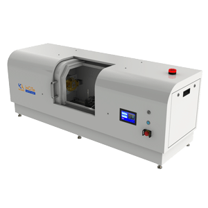
inCiTe™ 3D X-ray Microscope
High-resolution phase contrast micro-CT for laboratory-based structural analysis.
inCiTe™ 2.0
Integrated spectral and phase contrast micro-CT for multi-material research
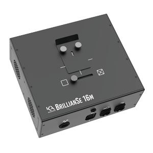
BrillianSe™ Detector
Direct conversion a-Se detector for high-resolution and low-flux research imaging.
Core Imaging Capabilities
Direct Conversion High-Resolution Detection
Direct conversion detection enables:
- High spatial resolution imaging
- Efficient performance above 20 keV
- Low-flux imaging efficiency
- Narrow point spread function (PSF)
- Micro-nano CT compatibility
Propagation-Based Phase Contrast Imaging
Phase contrast imaging enables:
- Improved visualization of low-density materials
- Enhanced edge definition
- Improved detectability of weakly absorbing structures
- Complementary contrast to absorption-based imaging
Spectral Multi-Energy Imaging
Scientific Research Applications
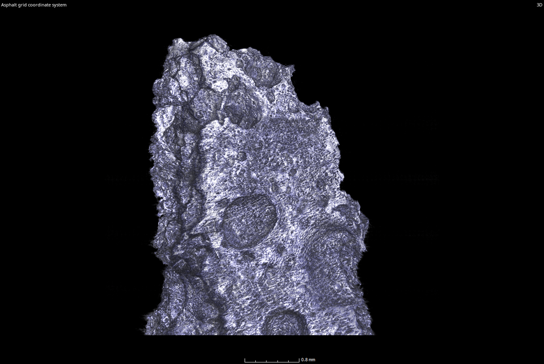
Materials Science and Composite Research
- Fiber composite analysis
- Additive manufacturing research
- Porosity and inclusion visualization
- Diffraction-based material microstructure imaging
- Internal structure characterization
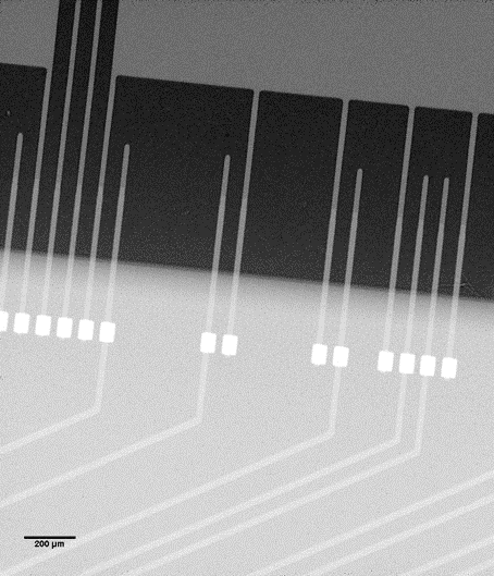
Semiconductor and Microelectronics R&D
- Device failure analysis
- Fine pitch trace inspection
- Crack and boundary visualization
- Laboratory-based microstructural analysis
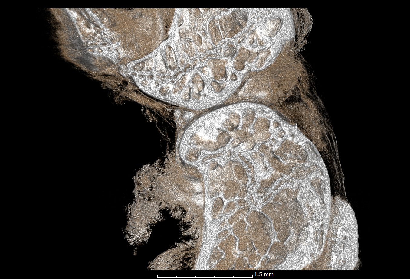
Biomedical and Preclinical Research Imaging
- Bone microstructure research
- Implant integrity analysis
- Soft tissue contrast enhancement (ex vivo)
- Pharmaceutical formulation inspection
- Specimen radiography
Propagation-based phase contrast imaging improves sensitivity to low-density and soft-tissue structures in laboratory and preclinical research environments.
Energy and Battery Materials Research
- Electrode morphology analysis
- Solid-state battery materials research
- Structural degradation studies
- Multi-material component differentiation
Why Multi-Modal X-ray Imaging Matters in Research
By integrating these modalities into laboratory-based systems, researchers can:
- Characterize complex multi-material samples
- Reduce destructive testing
- Improve microstructural analysis
- Enhance experimental reproducibility
- Streamline imaging workflows
Trusted by
Advanced Research Facilities and National Laboratories
BrillianSe™ direct conversion detectors are utilized in laboratory-based research environments requiring micron-scale resolution and efficient detection at elevated X-ray energies.
Research System Specifications
inCiTe™ 3D X-ray Microscope
- 8 µm pixel resolution
- Large 32 mm × 32 mm field of view
- Efficient low-flux imaging
- Grating-less phase contrast implementation
inCiTe™ 2.0 3D X-ray Microscope
An advanced modular micro-CT system integrating:
- Phase contrast imaging
- Spectral multi-energy imaging
- X-ray source options up to 130 kV
- Sub-micron pixel size at maximum magnification
- Larger sample accommodation via Reveal™ flat-panel integration
BrillianSe™ Direct Conversion Detector
A 16-megapixel hybrid a-Se/CMOS detector enabling:
- 8 µm pixel dimensions
- High DQE at hard X-ray energies
- Low-flux efficiency
- Diffraction-based microstructure imaging
- Micro-nano CT applications
Discuss Your Research Application
Latest blogs
What Is Low Flux Imaging? Low flux imaging refers to imaging conditions where only a…
You can’t fix what you can’t see. As electronic devices become more compact, densely layered,…
EOD teams depend on the accuracy of explosive detection equipment for keeping high-risk areas like…