
inCiTe™ Benchtop 3D X-ray Microscope
Benchtop micro-CT with phase contrast for superior clarity in low-density materials.
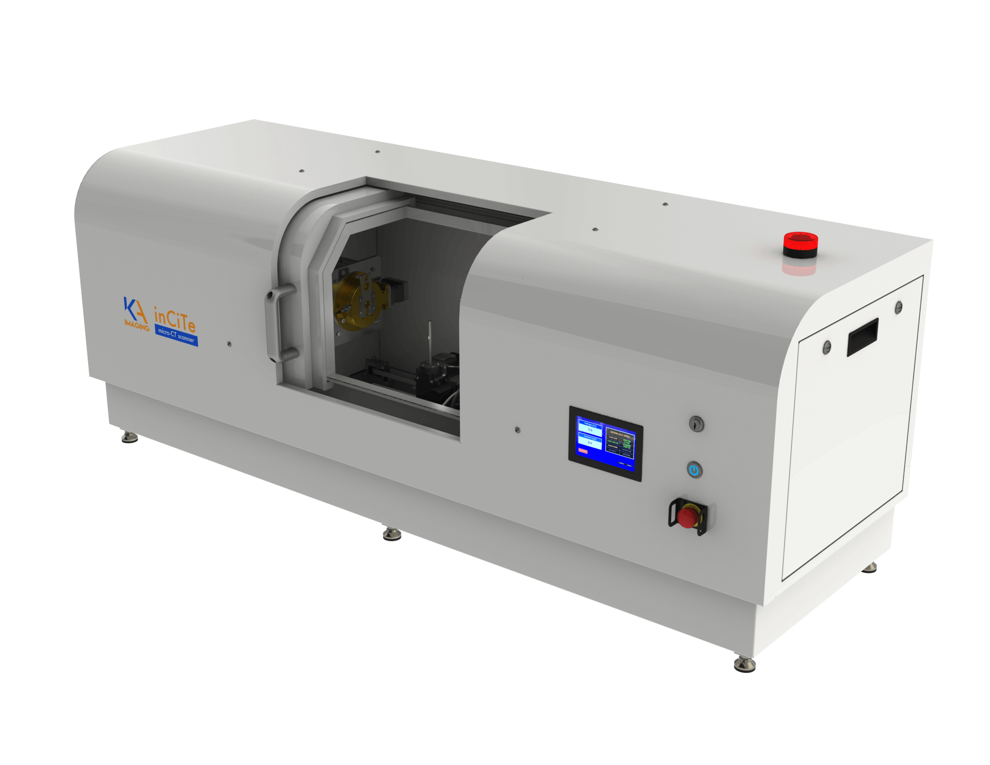
inCiTe™ Benchtop 3D X-ray Microscope for Phase-Contrast Imaging
The inCiTe™ 3D X-ray Microscope is a benchtop micro-CT system designed to enhance the visualization of low-density and weakly absorbing materials through advanced phase-contrast imaging.
By improving sensitivity to subtle structural variations, inCiTe enables detailed imaging of soft biological tissues, polymers, and other challenging samples that are difficult to assess with conventional absorption-based X-ray methods. Its compact design supports high-resolution, three-dimensional imaging across research and industrial environments.
inCiTe™ is the first commercial CT system to incorporate the BrillianSe™ amorphous selenium (a-Se) detector, combining high spatial resolution with efficient X-ray detection for precise imaging performance.
TECHNICAL SPECIFICATIONS
X-ray source
| Type | Maintenance-free, sealed |
|---|---|
| Smallest spot size | 5 µm (at 4 W) |
| Voltage | 40–100 kV |
| Maximum power | 20 W |
Imaging performance
| Spatial resolution | 11 µm 0.5 MTF at 45 cycles/mm5.6 µm 0.1 MTF at 90 cycles/mm |
|---|---|
| Pixel size at maximum magnification | 0.8 µm |
| X-ray phase contrast | Propagation-based |
| Image format | Raw (no header), 16-bit unsigned, little endian |
Detector
| Detector type | Selenium-CMOS direct conversion |
|---|---|
| Detector format | 16 MP (4k × 4k pixels) |
| Detector area | 32 × 32 mm² |
| Pixel pitch | 8 µm |
Sample and scanning geometry
| Maximum sample / scanning diameter | 25 mm |
|---|---|
| Maximum scanning length | 30 mm |
| Maximum sample length | 100 mm |
| Source-to-object distance | 44–410 mm |
| Source-to-detector distance | 84–450 mm |
| Lateral object translation | 50 mm |
| Vertical object translation | 12.7 mm |
| Rotational stepping | 0.01° (minimum) |
System, safety and installation
| Radiation safety | < 4 µSv/hr at 50 mm from any accessible surface |
|---|---|
| Dimensions (W × D × H) | 150 × 48 × 60 cm |
| Weight | 280 kg |
| Installation requirements | 120 V AC power 10–30 °C operating temperature < 85% humidity Zero condensation |
Product overview
X-ray detector: 16MP CMOS Detector, 8μm pixel, 14 bit resolution
Spatial Resolution: 11 µm ( 0.5 MTF at 45 cycles/mm); 5.6 μm ( 0.1 MTF at 90 cycles/mm
X-ray Source: 40-110 KV, 16 W, 2 μm spot size
Propagation-based X-ray phase contrast
Better visualization of low-density materials
Faster scan time
Literature
inCiTe™: Advancing 3D X-ray Imaging
inCiTe™, our cutting-edge 3D X-ray microscope, sets a new standard in scientific imaging and quality control. Powered by the innovative BrillianSe™ X-ray Detector, inCiTe™ delivers exceptional clarity and precision. Applications include:

Non-Destructive Testing (NDT)
Additive Manufacturing
Electronics
Agriculture

Geology
Preclinical Imaging
How an inCiTe scan works
- Step 1: Mount and position the sample.
- Step 2: Optimize the scan parameters and imaging geometry.
- Step 3: Acquire X-ray projections through sample rotation.
- Step 4: Reconstruct the 3D volume.
- Step 5: Visualize and analyze the results.
Customer Stories
What is Phase Contrast Imaging?
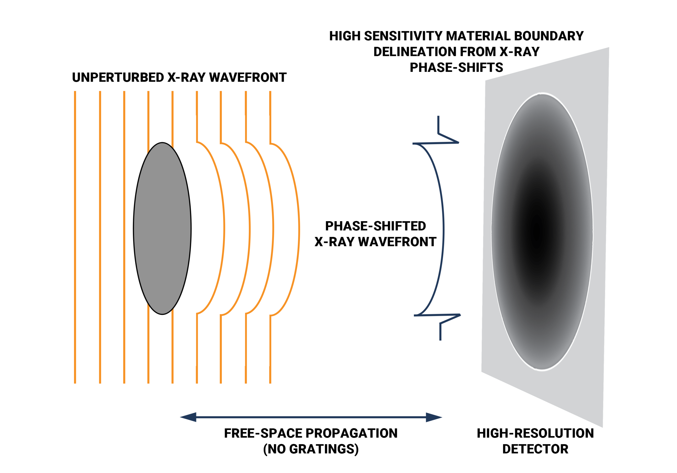
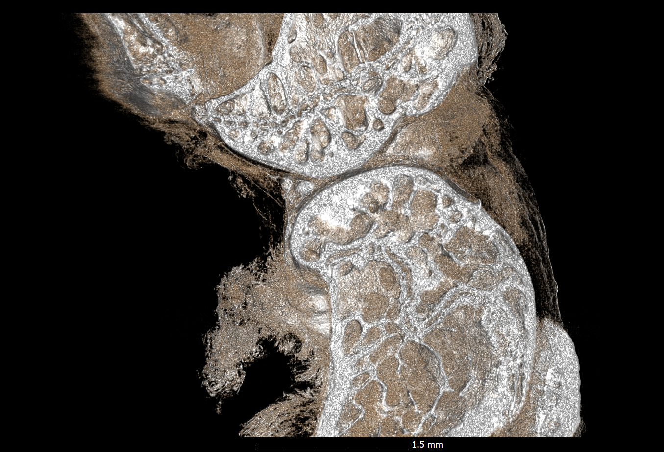
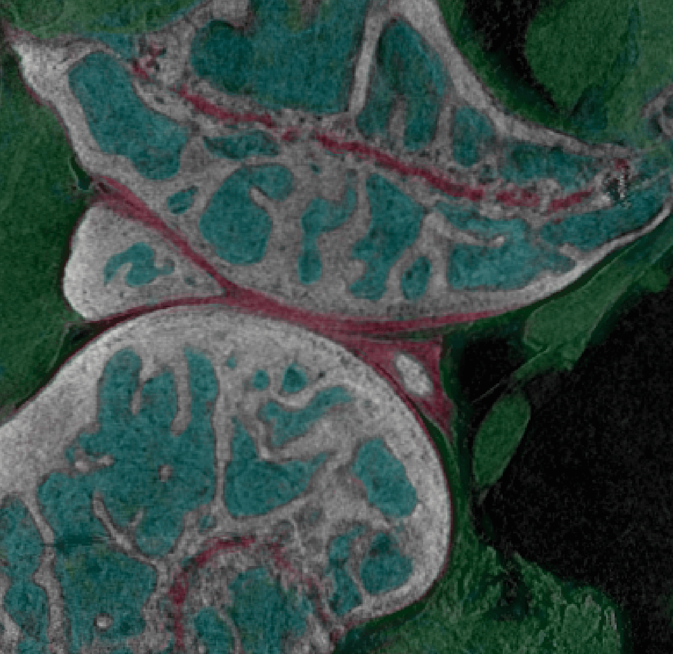
The inCiTe™ 3D X-ray Microscope excels in phase-contrast imaging, which is crucial when traditional X-ray methods struggle with low-density materials like soft tissues or polymers. Unlike conventional systems that measure only X-ray intensity, thanks to BrillianSe™ X-ray Detector, inCiTe™ can capture subtle phase shifts, enhancing image contrast. This is achieved using free-space propagation, where phase variations translate directly into intensity fluctuations, offering clearer, more detailed images.
How BrillianSe™ Works
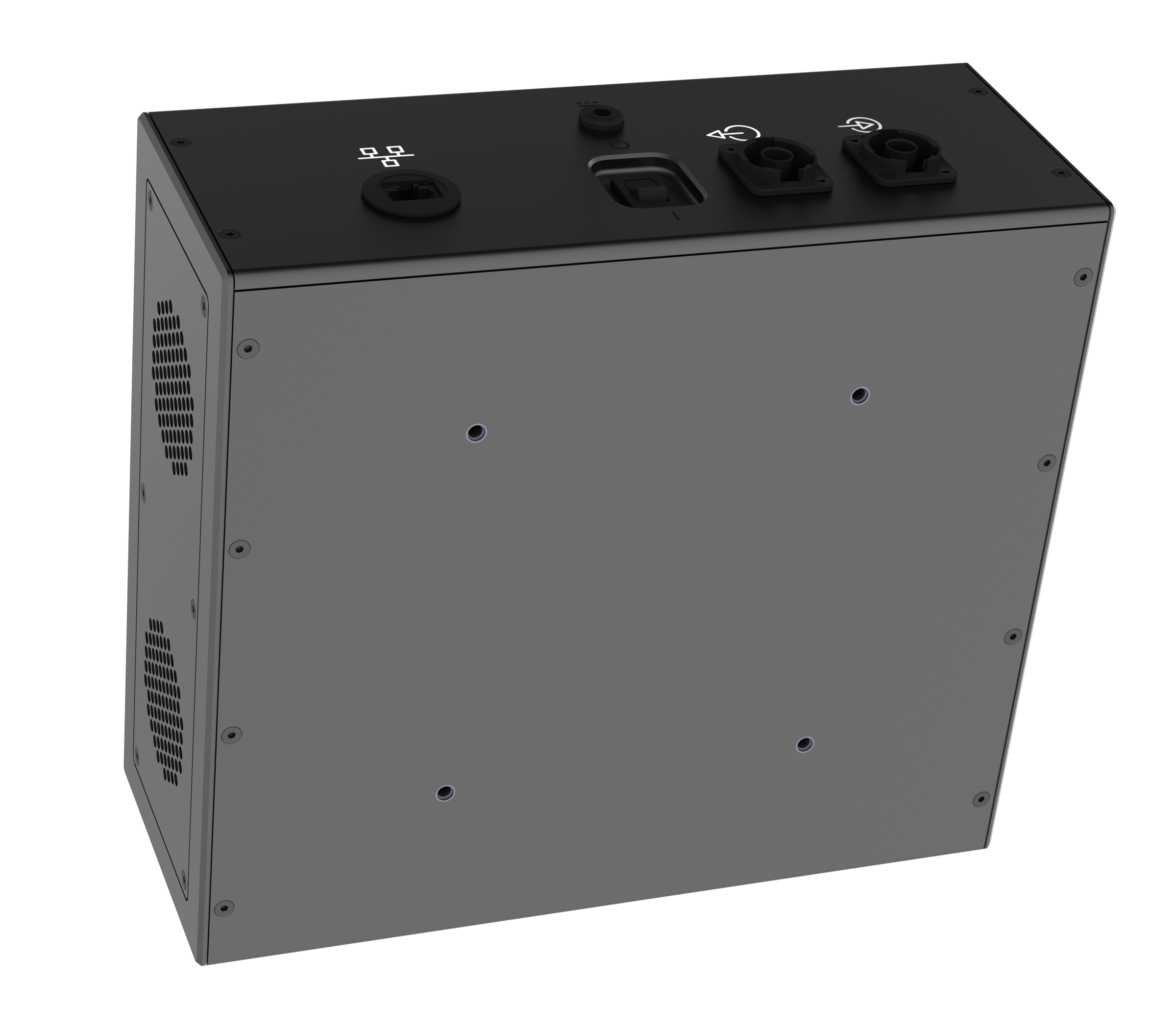
BrillianSe™ is a hybrid a-Se/CMOS detector that uses an a-Se photoconductor with high intrinsic spatial resolution for direct conversion of X-ray photons to electric charge. The electronic signal is then read out by a low noise CMOS active pixel sensor (APS). Without the need to first convert X-ray photons to visible light, as in indirect scintillator-based approaches, thinning of the conversion layer to minimize optical scatter is not necessary.
Looking for Advanced Material Differentiation? Explore inCiTe™ 2.0
The inCiTe™ 3D X-ray Microscope is designed to deliver high-resolution phase-contrast imaging for detailed visualization of low-density and weakly absorbing materials.
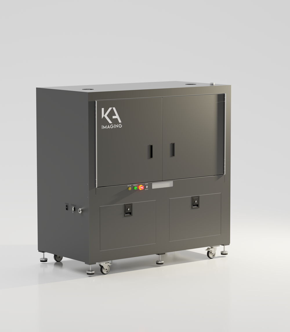
For applications requiring additional material differentiation, inCiTe™ 2.0 builds on this foundation by integrating spectral imaging capabilities alongside phase contrast. This enables the analysis of complex and multi-material samples within a single scan, extending beyond structural imaging to provide additional insight into material composition.
Explore inCiTe™ 2.0, our Spectral 3D X-ray Microscope:
Which one is for me? inCiTe vs. inCITe 2.0
While both systems utilize our patented phase-contrast technology, they are designed for different operational environments. Paired with the BrillianSe detector, the inCiTe is our dedicated benchtop specialist, offering a compact, space-efficient footprint for researchers focused on high-resolution imaging of low-density materials (up to 110 kV).
In contrast, the inCiTe 2.0 is an expanded research workstation that can be configured with either the BrillianSe or Reveal detectors. It is built to accommodate high-power 150 kV sources and provides the expanded internal cabinet volume necessary for custom in situ stages and complex biomechanical rigs.
|
Feature / Capability
|
inCiTe (Phase Contrast)
|
inCiTe 2.0 (Spectral)
|
|---|---|---|
|
Dimensions |
Dedicated Phase Contrast clarity in a small footprint. |
Spectral imaging + high-load In Situ flexibility. |
|
Max X-ray Source |
110 kV |
Up to 150 kV |
|
Primary Technology |
Propagation-based Phase Contrast |
Phase Contrast + Spectral (Color) Imaging |
|
Material Specialty |
Low-Density Materials: |
Multi-Material Discovery: |
|
Key Advantage |
Maximum contrast for nearly "invisible" soft samples. |
Ability to "color-code" materials based on atomic number (Z). |
|
Imaging Modes |
Absorption and Phase Contrast. |
Absorption, Phase Contrast, and Spectral Material Decomposition. |
|
Sample Density |
Optimized for low to medium-density samples. |
High versatility; handles low-density to high-density industrial parts. |
|
Best For... |
Analytical Researchers: |
Biomechanical & Industrial Engineers: |
Is a Benchtop Micro-CT Right for Your Lab?
inCiTe™ is built for labs that need micrometer-scale 3D imaging from a compact benchtop system, particularly for low-density and weakly absorbing samples such as soft tissue, polymers, foams and fibers that can be difficult to visualize with conventional absorption micro-CT. With a system in your own lab, researchers can run scans around their own workflows rather than relying exclusively on shared imaging facilities or outsourced analysis.
For larger, denser or multi-material samples, inCiTe™ 2.0 expands the platform with spectral imaging, a higher-energy X-ray source and greater sample capacity.
Not sure which system is the right fit? Our team is happy to talk through your samples and imaging goals to help identify the best option.
Media Gallery
Other products that might interest you
Latest blogs
Beyond BMD: Understanding Bone Structure Two treatment groups in a preclinical study may show similar…
Looking Beyond the Surface You’ve inspected the surface of a failed component and found nothing…
Biomedical research often requires scientists to visualize structures with vastly different X-ray attenuation properties within…
Original research
S. Abbaszadeh, CC Scott, O Bubon, A Reznik, KS Karim, “Enhanced detection efficiency of direct conversion X-ray detector using polyimide as hole-blocking layer,” Scientific Reports, December 2013
G.P. Lindberg, T. O’Loughlin, N. Gross, A. Mishchenko, A. Reznik, S. Abbaszadeh, K.S. Karim, G. Belev, B. Weinstein, “Photo-crystallization in a-Se layer structures: effects of film-substrate interface-rigidity,” Journal of Applied Physics, vol. 116, November 2014.
A. Parsafar, C. Scott, A. El-Falou, P. Levine, K.S. Karim, “Direct-Conversion CMOS X-Ray Imager with 5.6 μm × 6.25 μm Pixels,” IEEE Electron Device Letters, 36(5), pp. 481-3, May 2015.
C.C. Scott, A. Parsafar, A. El-Falou, P. M. Levine, K.S. Karim, “High Dose Efficiency, Ultra-high Resolution Amorphous Selenium/CMOS Hybrid Digital X-ray Imager,” IEEE International Electron Devices Meeting (IEDM) Technical Digest, December 2015.
Procházková, P., Kaiser, M., Stravová, Z., Petřík, M., Karim, K. S., Birch, Z., Comai, G. E., Corb, A., Tajbakhsh, S., Zikmund, T., & Kaiser, J. (2025, September). Revealing the architecture of eye muscles using cryogenic X-ray imaging [Oral presentation]. Tomography for Scientific Advancement (ToScA) Conference, France.
Related research
- Comparing conventional and phase contrast imaging
Gureyev, Timur Eugenievich, Sheridan C. Mayo, Damian E. Myers, Ya Nesterets, D. M. Paganin, A. Pogany, Andrew W. Stevenson, and S. W. Wilkins. “Refracting Röntgen’s rays: propagation-based x-ray phase contrast for biomedical imaging.” Journal of Applied Physics 105, no. 10 (2009): 102005.
Krenkel, Martin, Mareike Töpperwien, Christian Dullin, Frauke Alves, and Tim Salditt. “Propagation-based phase-contrast tomography for high-resolution lung imaging with laboratory sources.” AIP Advances 6, no. 3 (2016): 035007.
Olivo, A., and E. Castelli. “X-ray phase contrast imaging: From synchrotrons to conventional sources.” La Rivista Del Nuovo Cimento 37 (2014): 467-508.
Bravin, Alberto, Paola Coan, and Pekka Suortti. “X-ray phase-contrast imaging: from pre-clinical applications towards clinics.” Physics in Medicine & Biology 58, no. 1 (2012): R1.
Bidola, P., K. Morgan, M. Willner, A. Fehringer, S. Allner, F. Prade, F. Pfeiffer, and K. Achterhold. “Application of sensitive, high‐resolution imaging at a commercial lab‐based X‐ray micro‐CT system using propagation‐based phase retrieval.” Journal of Microscopy 266, no. 2 (2017): 211-220.
- Propagation
Wilkins, S. W., T. Ei Gureyev, D. Gao, A. Pogany, and A. W. Stevenson. “Phase-contrast imaging using polychromatic hard X-rays.” Nature 384, no. 6607 (1996): 335-338.
Mayo, Sheridan C., Andrew W. Stevenson, and Stephen W. Wilkins. “In-line phase-contrast X-ray imaging and tomography for materials science.” Materials 5, no. 5 (2012): 937-965.
- Gratings
Pfeiffer, Franz, Timm Weitkamp, Oliver Bunk, and Christian David. “Phase retrieval and differential phase-contrast imaging with low-brilliance X-ray sources.” Nature physics 2, no. 4 (2006): 258-261.
Frequently Asked Questions
What is a Micro-CT?
Micro-CT (micro-computed tomography) is an X-ray imaging technique that produces 3D images of an object’s internal structure at micrometer-scale resolution. The sample rotates while a series of 2D X-ray images are captured from different angles. Software then reconstructs these images into a 3D volume that can be viewed slice by slice in any direction, rendered in 3D, and measured, typically without cutting the sample open. Micro-CT is widely used in materials science, manufacturing quality control, geology, and biomedical research.
What is a 3D X-ray Microscope?
A 3D X-ray microscope (XRM) is a high-resolution X-ray imaging system that reveals the internal 3D structure of small samples without sectioning them. Like micro-CT, it captures X-ray images as the sample rotates and reconstructs them into a 3D volume. The inCiTe™ is a benchtop 3D X-ray microscope with propagation-based phase contrast, which improves the visibility of low-density materials such as soft tissue, polymers, foams, and fibers.
Is micro-CT non-destructive?
Yes. Micro-CT uses X-rays to image the inside of a sample, so there’s no need to cut, section, or polish it. After scanning, the same sample can be rescanned, tested by other methods, or kept intact. This makes micro-CT well suited to failure analysis, quality control, and rare or one-of-a-kind specimens. Some samples are sensitive to radiation, such as living specimens and certain biological or polymer materials, so scan settings should be chosen with the sample in mind.
What resolution can inCiTe reach?
inCiTe™ achieves a spatial resolution of 11 µm (at 0.5 MTF) and 5.6 µm (at 0.1 MTF) using its 8 µm pixel BrillianSe™ detector. Spatial resolution reflects the smallest features the system can actually distinguish, which makes it a more meaningful measure of image detail than pixel or voxel size alone. The resolution achieved on a given scan also depends on sample size and scan settings.
How long does a scan take?
Many inCiTe™ scans can be completed in under 20 minutes with minimal setup. Actual scan time depends on the sample’s size and density, the resolution required, the number of projections collected, and the imaging mode. Quick overview scans take less time, and high-resolution scans of fine detail take longer.
Do soft-tissue samples need staining?
Not always. Soft tissue absorbs X-rays weakly, so conventional absorption micro-CT often relies on iodine- or heavy-metal-based stains to increase contrast. Staining adds preparation time and can alter the specimen, depending on the stain and protocol.
inCiTe™ uses propagation-based phase contrast, which is sensitive to changes in X-ray phase as well as absorption. This can improve the visibility of low-density and weakly absorbing structures and enable imaging without contrast staining.
What sample sizes can inCiTe image?
inCiTe™ is designed for small samples, with a maximum object size of 32 mm in its largest dimension. Smaller samples can be positioned for greater geometric magnification, which reduces effective pixel size and can help visualize finer features. The optimal setup depends on the sample size, region of interest, and imaging task.
For larger samples, inCiTe™ 2.0 offers a substantially larger sample capacity.