UPGRADE TO REVEAL 35C: AN ECONOMIC PROPOSITION
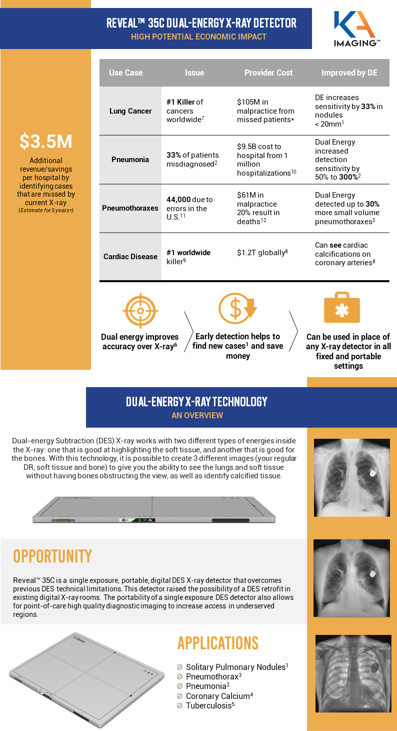
Dual energy has been shown to detect more solitary pulmonary nodules, pneumothorax, pneumonia, tuberculosis and coronary calcium than conventional analog or digital chest X-ray. This early detection enables better patient outcomes, additional hospital revenue, cost savings to healthcare systems and reduces exposure to malpractice claims.
KA Imaging has created Reveal™ 35C, the world’s first digital portable dual-energy X-ray detector as a universal upgrade for any analog or digital X-ray detector from any manufacturer.
The infographic below summarizes the study.

UPGRADE TO REVEAL 35c: AN ECONOMIC PROPOSITION
Fill the form to receive the complete study in your email.
References
1.Oda, Seitaro, Kazuo Awai, Yoshinori Funama, Daisuke Utsunomiya, Yumi Yanaga, Koichi Kawanaka, Takeshi Nakaura et al. “Detection of small pulmonary nodules on chest radiographs: efficacy of dual-energy subtraction technique using flat-panel detector chest radiography.” Clinical radiology 65, no. 8 (2010): 609-615.
2.Martini, Katharina, Marco Baessler, Stephan Baumueller, and Thomas Frauenfelder. “Diagnostic accuracy and added value of dual-energy subtraction radiography compared to standard conventional radiography using computed tomography as standard of reference.” PloS one 12, no. 3 (2017): e0174285.
3.Urbaneja, A., Dodin, G., Hoosu, G., et al. (2018) Added Value of Bone Subtraction in Dual-energy Digital Radiography in the Detection of Pneuomothorax: Impact of Reader Expertise and Medical Specialty. The Association of University Radiologists. Elsevier Inc.
4. Song, Yingnan, Hao Wu, Di Wen, Bo Zhu, Philipp Graner, Leslie Ciancibello, Haran Rajeswaran et al. “Detection of coronary calcifications with dual energy chest X-rays: clinical evaluation.” The International Journal of Cardiovascular Imaging (2020): 1-8.
5.Sharma, Madhurima, Manavjit Singh Sandhu, Ujjwal Gorsi, Dheeraj Gupta, and Niranjan Khandelwal. “Role of digital tomosynthesis and dual energy subtraction digital radiography in detection of parenchymal lesions in active pulmonary tuberculosis.” European Journal of Radiology 84, no. 9 (2015): 1820-1827.
6.Kuhlman, Janet E., Jannette Collins, Gregory N. Brooks, Donald R. Yandow, and Lynn S. Broderick. “Dual-energy subtraction chest radiography: what to look for beyond calcified nodules.” Radiographics 26, no. 1 (2006): 79-92.
7.https://www.who.int/news-room/fact-sheets/detail/cancer
8.Song, Yingnan, Hao Wu, Di Wen, Bo Zhu, Philipp Graner, Leslie Ciancibello, Haran Rajeswaran et al. “Detection of coronary calcifications with dual energy chest X-rays: clinical evaluation.” The International Journal of Cardiovascular Imaging (2020): 1-8.
9.https://www.who.int/health-topics/cardiovascular-diseases#tab=tab_1
10.Divino, V., etal “The annual economic burden among patients hospitalized for community-acquired pneumonia (CAP): a retrospective US cohort study.” Current Medical Research and Opinion 36, no. 1 (2020): 151-160.
11.https://www.healthgrades.com/quality/patient-safety-2020-infographic
12.Domino etal, “Injuries and Liability Related to Central Vascular Catheters–A Closed Claims Analysis”, Anesthesiology 2004; 100:1411-8.
* Contact us to see the full study
