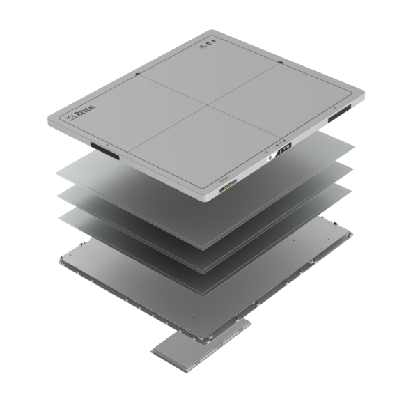
Reveal™ 35C has a unique triple stacked layer design, allowing for easy integration and high quantum efficiency. The detector works with a single X-ray exposure, eliminating motion artifacts. This ground-breaking Dual-Energy Subtraction (DES) technology is called SpectralDR™. With KA Imaging’s patented SpectralDR™ technology, the full spectrum and multiple energies are acquired in one standard chest X-ray exposure, thus maintaining the radiation dose of a conventional chest X-ray. Our technology produces sharp X-ray images that contain zero motion artifacts.
SpectralDR™ offers improved visualization of lung nodules, pneumonia, line and tube tips, pneumothorax, retained surgical objects and more. This 3-in-1 solution simultaneously acquires 3 images in 1 single exposure, improving visualization of bone and tissue.
REVEAL™ 35C
Revealᵀᴹ 35C is the world’s first single exposure dual-energy X-ray detector that can be used in both fixed and mobile/portable applications. Unlike other dual-energy solutions, Reveal uses the same radiation dose as a regular chest X-ray, allowing for lateral dual-energy images for the first time.
Single Exposure
Fixed and mobile/portable applications
Same radiation dose as a chest X-ray
Multiple Views: PA, Lateral, Oblique
Exclusive Technolgy
CLINICAL APPLICATIONS
Pulmonary Nodules
Pneumonia
Pneumothorax
Coronary Calcium
PICC lines
Intravenous pyelogram (IVP)
Pneumoconiosis (black lung)
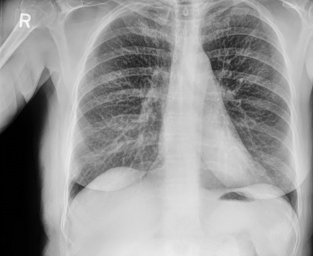
IMAGE GALLERY
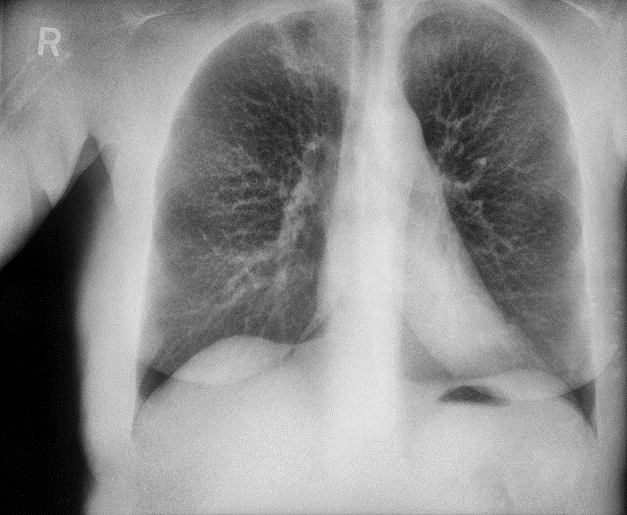
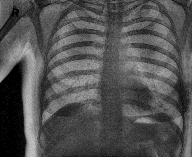
Hidden Apical Lesion in RUL
Apical lesion missed in Conventional due to overlapping bone. Old rib fracture also became visible on right side.
DR
Soft tissue image
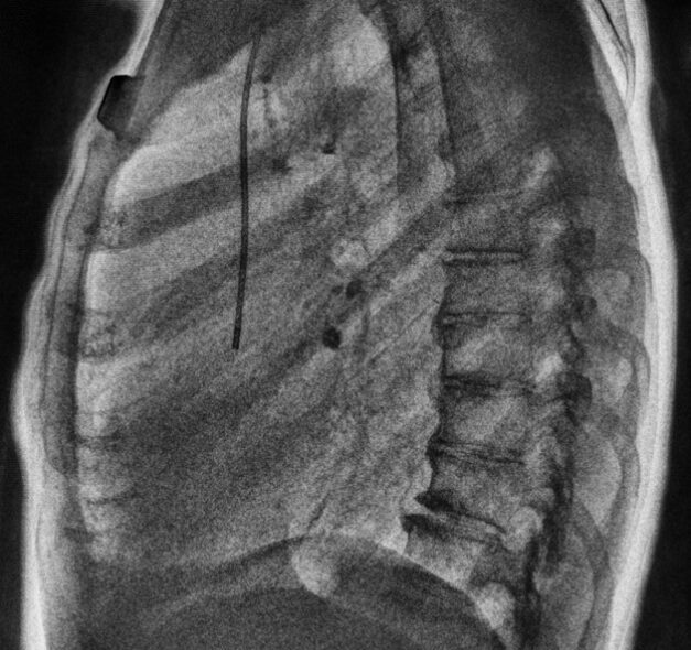
Bone Image
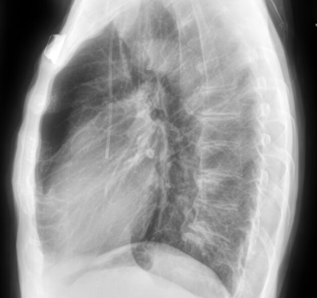
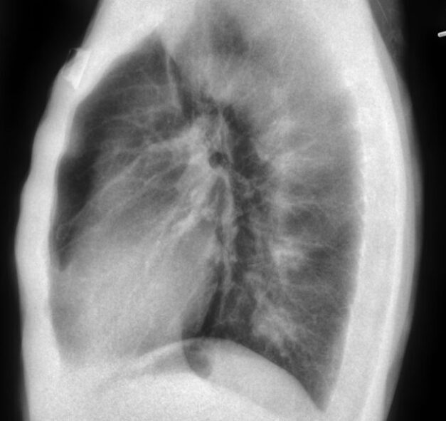
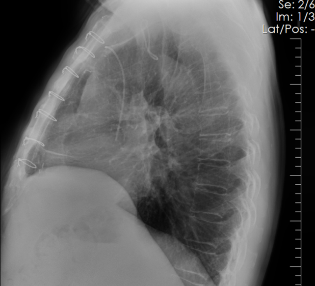
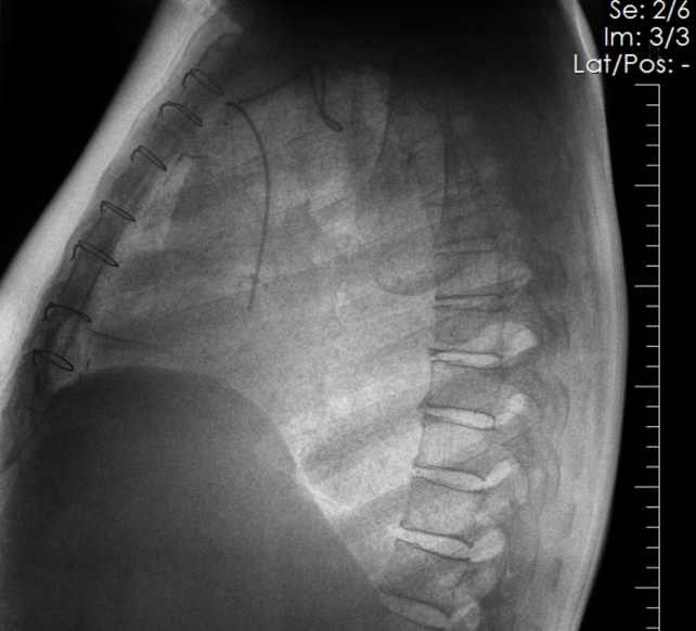
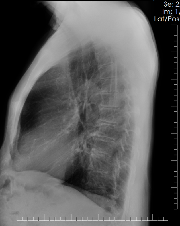
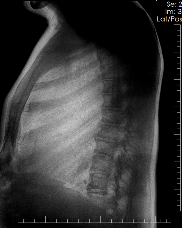
Hidden Mass in RLL Obscured by Degenerative Bone Disease (Lateral)
Mass seen behind heart superimposed on calcified joint. Clearly seen portocath line and sternum in bone image.
DR
Soft tissue image
Bone Image
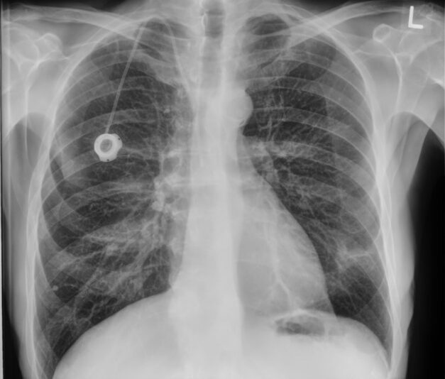
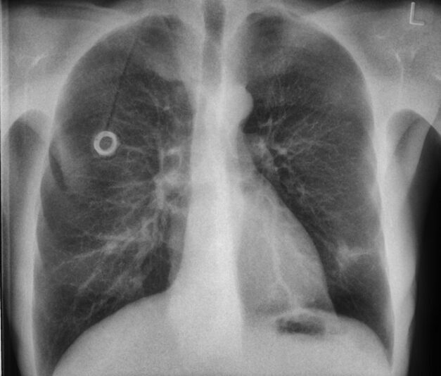
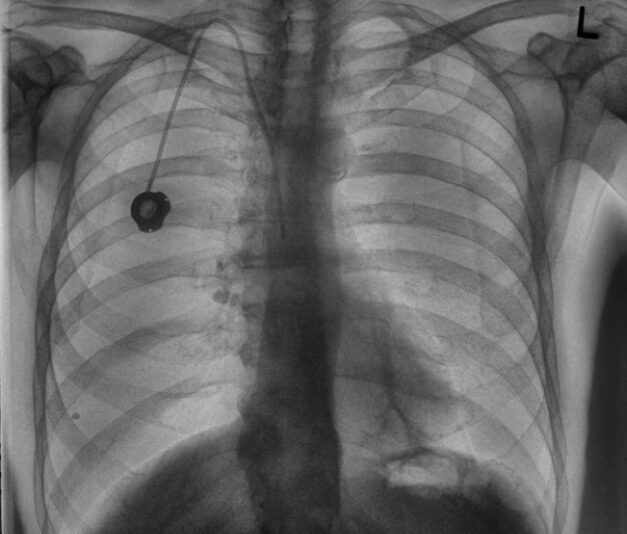
Hidden Mass in RLL Obscured by Degenerative Bone Disease (PA)
Mass behind heart is not seen in PA image. Line easier to see on Bone image.
DR
Soft tissue image
Bone Image
Coronary Calcium (Lateral)
Calcified coronary seen in lateral image. Clearly seen portocath line and sternum in bone image.
DR
Soft tissue image
Bone Image
Coronary Stents
Cardiac calcifications and Stents seen clearly using a lateral image