
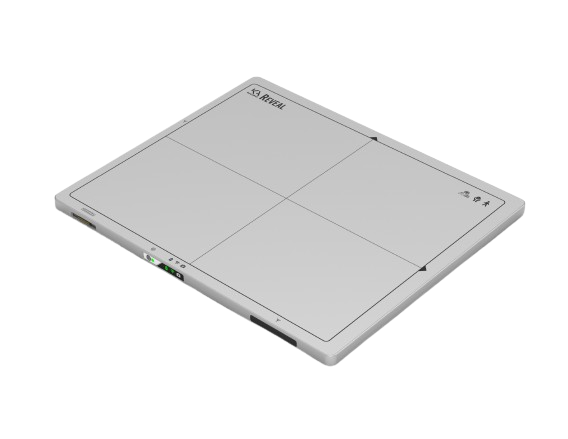
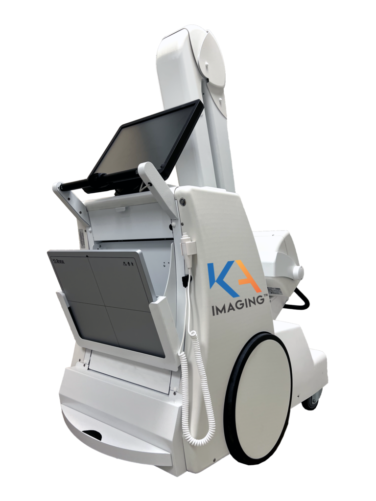
Reveal™ 35C X-ray Detector
Upgrade Your Existing X-ray System with SpectralDR® Dual-Energy Imaging
Reveal™ 35C is the first and only single-exposure SpectralDR® detector, delivering dual-energy images — standard DR, bone, and soft tissue — without the workflow disruptions or limitations of traditional dual-energy systems. With a single exposure, free from motion artifacts, Reveal™ 35C can perform any X-ray exam or positioning requirement, upgrading your imaging capabilities without adding dose or complexity.
Not in the right place? Explore this product in NDT | Veterinary
More Insight from Every X-ray
Reveal™ 35C works with your current protocols and workflow — then gives you the power to optimize based on your diagnostic needs.
Reveal 35C can be used for general radiography applications. Studies have shown benefits in assessing lung conditions such as pneumonia and pneumothorax, as well as in visualizing lung lesions and confirming line or tube placement — all without additional scans, dose, or hardware.
Independent researchers are also investigating emerging applications like the visualization of coronary calcium10,11,12.
Key Features
SpectralDR® technology:
1 exposure, 3 images, 0 motion artifacts
Cassette-size: 14 x 17 inch / 35 x 43 cm (ISO 4090) for seamless upgrade
Triple-layer detector design delivers a DQE of 67% at 1 lp/mm — up to 30% higher than typical DR panels.
True dual-energy subtraction through physics
Hot Swappable Batteries
Literature
Recent Clinical Findings
See published research, case studies, and technical briefs in our X-ray Academy (free account required).
Why choose dual-energy (Spectral) X-ray?
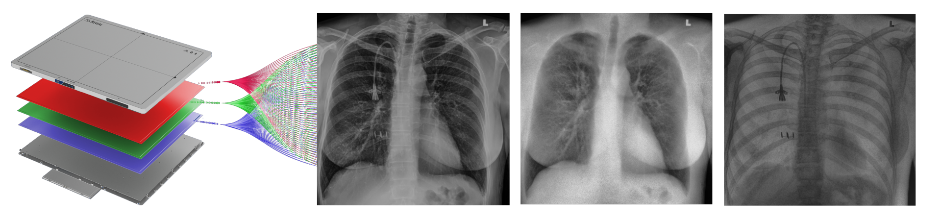
SpectralDR® technology enables simultaneous acquisition of:
Standard X-ray
Soft tissue image
Bone image
—all from a single exposure using a standard X-ray dose.
Dual-energy images are clinically proven1 to:
Improve visibility of lung nodules, pneumonia, pneumothorax, and calcifications
Provide clearer identification of lines, tubes, and retained surgical objects
Improve image clarity without increasing dose or scan time
Impact and ROI
See how integrating dual-energy mobile X-ray could help reduce downstream imaging costs, support earlier, more confident diagnoses, and improve resource utilization in critical care settings.
Clinical Case
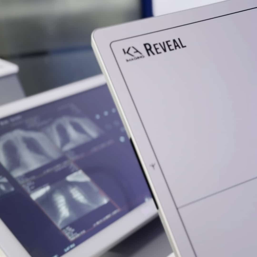
Reveal™ 35C with SpectralDR® technology brings dual-energy imaging to any compatible X-ray system. Whether in general radiography, emergency care, or critical care units, the ability to capture standard, bone, and soft tissue images in a single exposure works with your current protocols and workflow — then gives you the power to optimize based on your diagnostic needs.
Hidden Pneumonia Detection
A 51-year-old leukemia patient had a normal DR image. The soft tissue image revealed a focal opacity, later confirmed as pneumonia on CT, showing how dual-energy imaging can reveal findings standard X-ray may miss.
DR/SoftTissue Image


DR/ Soft Tissue Image
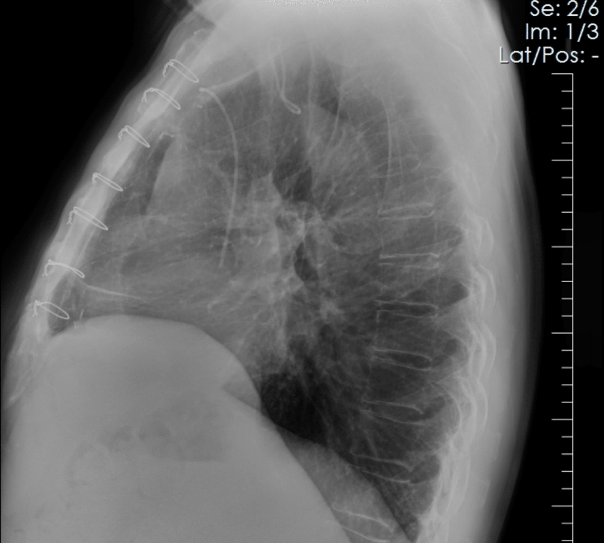
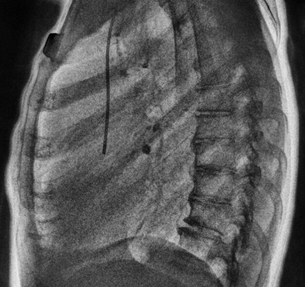
Visualization of calcifications
Lateral dual-energy bone image shows visible calcified coronary vessels, along with a PortoCath line, sternum, and sternal wire ties — all in a single exposure with Reveal™ 35C and SpectralDR® technology.
DR/Bone Image

Hidden mass Obscured by Degenerative Bone Disease
DR/SoftTissue Image


DR/ Soft Tissue Image
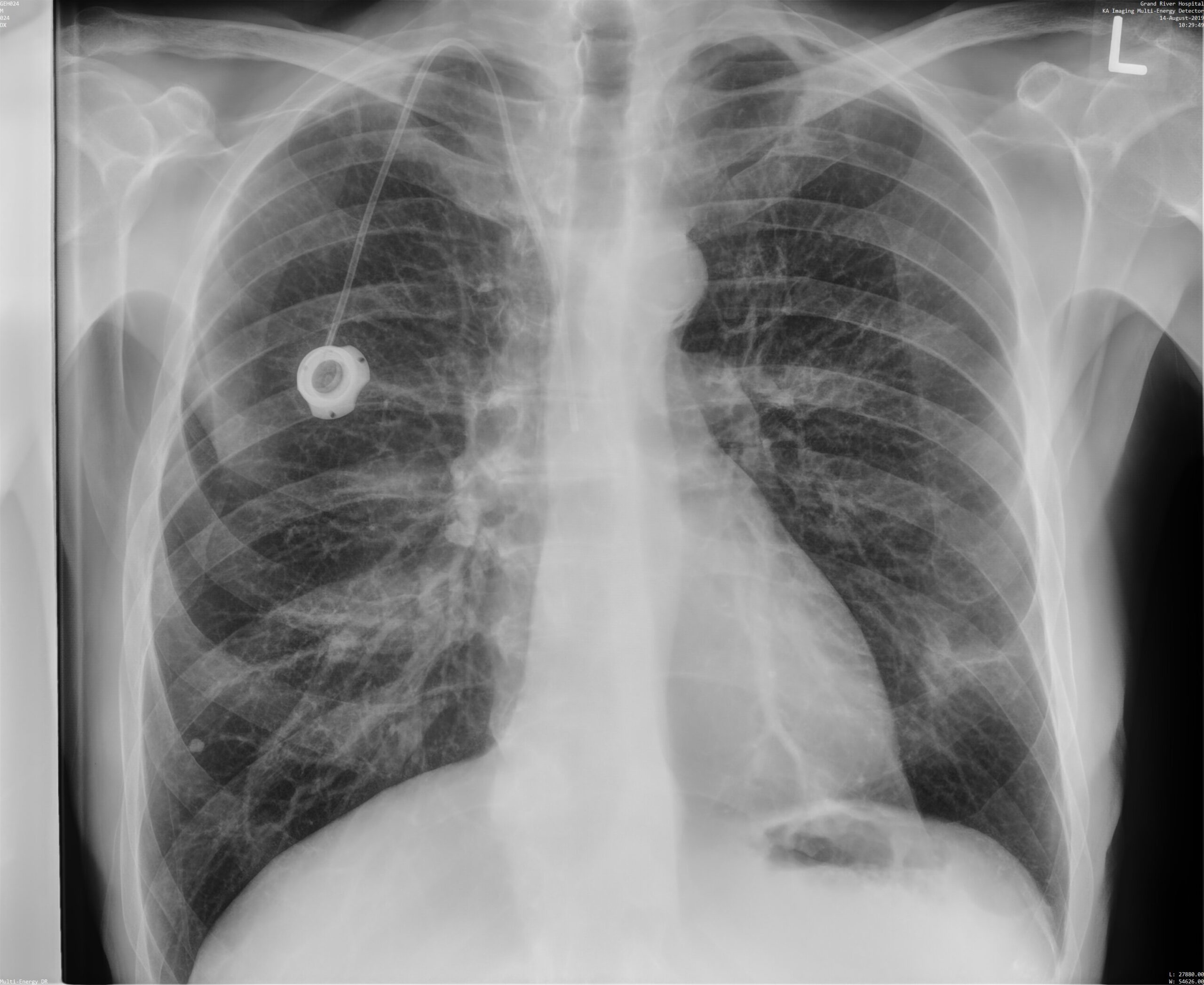
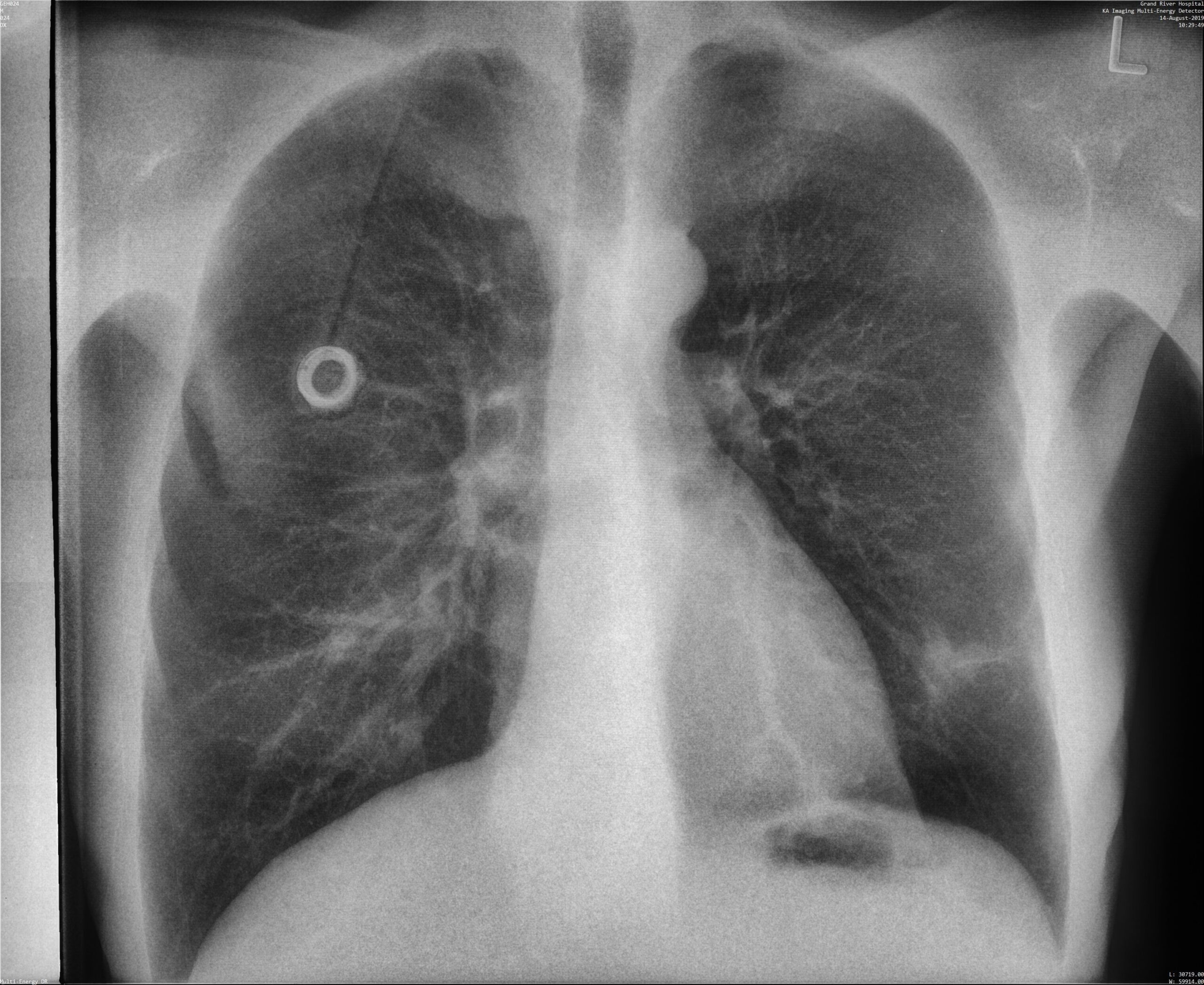
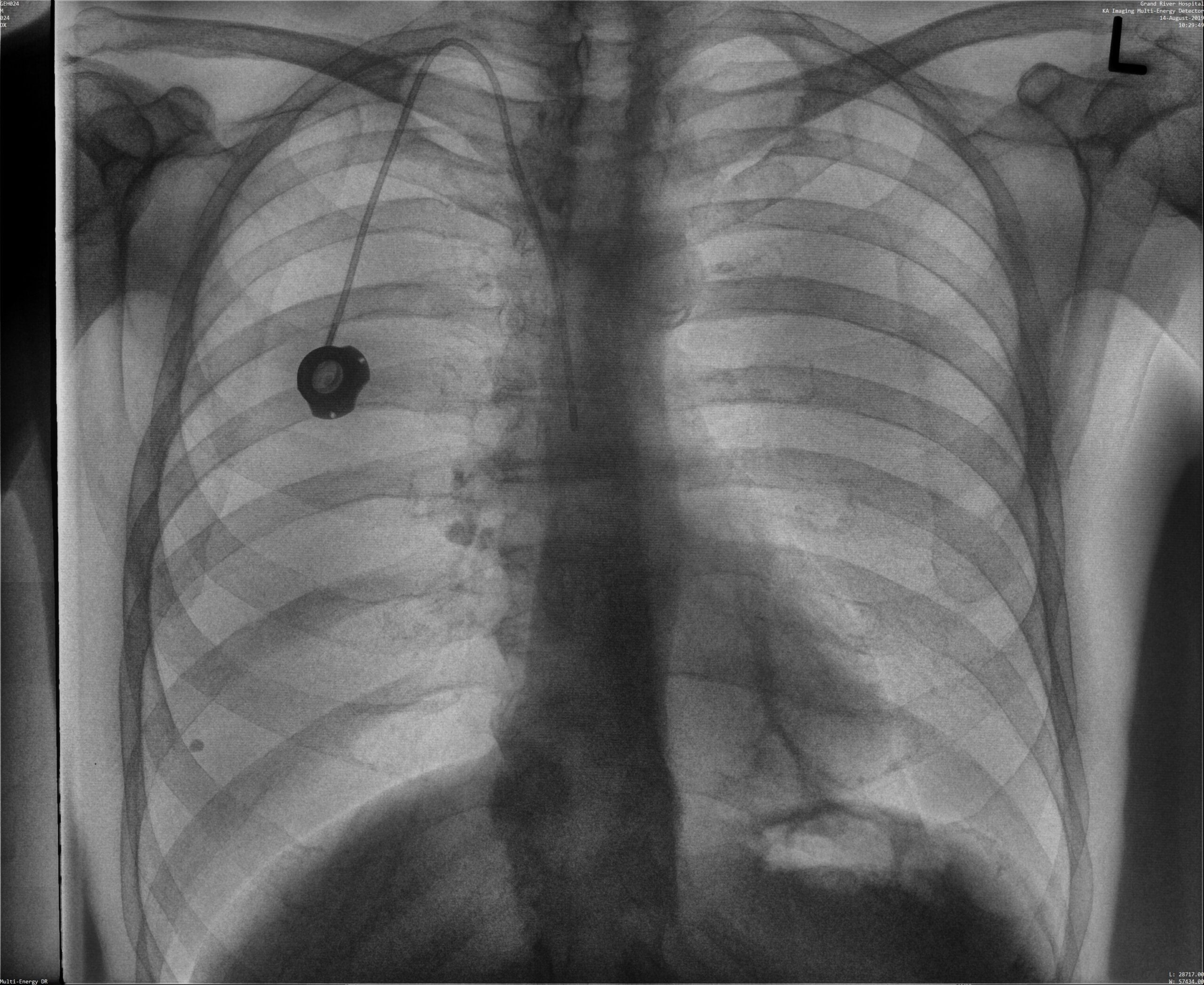
Better Line Placement Confirmation
Spectral dual-energy bone images provide clear visualization of vascular wire mesh and tube tips — without extra software, dose, or delay.
DR/Bone Image


Reveal™ 35C: Evidence from Clinical Studies

33% more pneumonia cases found
compared to X-ray thanks to dual-energy images⁴

Increased confidence in 57% of ICU cases
thanks to dual-energy images with no added reading time⁵

43% more lesion visibility
when dual-energy images were included³

Up to 37% decrease
in the number of follow up CT scans for ICU patients9

Better visibility of lines and tube tips
without extra reading times⁵
Reveal 35C intended use: The Reveal 35C Flat Panel Detector (FPD), when used with a radiographic imaging system, is intended to generate radiographic images of human anatomy wherever a conventional screen-film, digital radiography (DR), or computed radiography (CR) detector is used for general purposes. Reveal 35C is designed to retrofit into an existing X-ray system, and for integration into a complete X-ray system by a qualified system integrator. When the dual energy subtraction function is enabled, it is intended to assist the physician through the visualization of anomalies by reducing the visibility of underlying or overlying anatomical structures. This device is not intended for use in mammography applications.
Trusted by Users
Media Gallery
Latest blogs
Every budget season, hospitals ask imaging leaders to predict the future using yesterday’s tools. Capital…
Traditionally, X-ray technology has not been able to come to space, but the single exposure…
Dual-energy X-ray imaging has traditionally been associated with large, fixed radiography systems and complex workflows.…
FDA 510(k) Clearance
Health Canada Medical Device Licence
CE Marking Under EU MDR
Contact us for availability in your region.
References:
- Improved patient outcomes for dual-energy subtraction imaging:
1.1 (Lung Nodules) Oda, Seitaro, Kazuo Awai, Yoshinori Funama, Daisuke Utsunomiya, Yumi Yanaga, Koichi Kawanaka, Takeshi Nakaura et al. “Detection of small pulmonary nodules on chest radiographs: efficacy of dual-energy subtraction technique using flat-panel detector chest radiography.” Clinical radiology 65, no. 8 (2010): 609-615.
1.2 (Pneumothorax) Urbaneja, A., Dodin, G., Hoosu, G., et al. (2018) Added Value of Bone Subtraction in Dual-energy Digital Radiography in the Detection of Pneuomothorax: Impact of Reader Expertise and Medical Specialty. The Association of University Radiologists. Elsevier Inc.
1.3 (Pneumonia) Martini, Katharina, Marco Baessler, Stephan Baumueller, and Thomas Frauenfelder. “Diagnostic accuracy and added value of dual-energy subtraction radiography compared to standard conventional radiography using computed tomography as standard of reference.” PloS one 12, no. 3 (2017): e0174285.
1.4 (Tuberculosis) Sharma, Madhurima, Manavjit Singh Sandhu, Ujjwal Gorsi, Dheeraj Gupta, and Niranjan Khandelwal. “Role of digital tomosynthesis and dual energy subtraction digital radiography in detection of parenchymal lesions in active pulmonary tuberculosis.” European Journal of Radiology 84, no. 9 (2015): 1820-1827.
1.5 (Coronary Calcifications) Song, Yingnan, Hao Wu, Di Wen, Bo Zhu, Philipp Graner, Leslie Ciancibello, Haran Rajeswaran et al. “Detection of coronary calcifications with dual energy chest X-rays: clinical evaluation.” The International Journal of Cardiovascular Imaging (2020): 1-8.
- Maurino, S. L., Badano, A., Cunningham, I. A., & Karim, K. S. (2016, March). Theoretical and Monte Carlo optimization of a stacked three-layer flat-panel x-ray imager for applications in multi-spectral diagnostic medical imaging. In Medical Imaging 2016: Physics of Medical Imaging (Vol. 9783, p. 97833Z). International Society for Optics and Photonics.
- S. L. Maurino, K. S. Karim, V. Venkatesh. Diagnostic value of single-exposure dual-energy subtraction radiography in lung lesion detection: initial results. European Congress of Radiology-ECR 2022, 2022.
- Sanchez F, Kandel S, May M, Ronghe S, Rogalla P. Diagnostic value of dual-energy chest x-ray in immunocompromised patients to rule out pneumonia: initial results. European Congress of Radiology-ECR 2021, 2021.
- Rogalla P., Karim K. Added diagnostic value of portable dual-energy chest X-ray in a non-radiological reviewing environment. Radiological Society of North America-RSNA 2022, 2022
- Kuhlman, Janet E., Jannette Collins, Gregory N. Brooks, Donald R. Yandow, and Lynn S. Broderick. “Dual-energy subtraction chest radiography: what to look for beyond calcified nodules.” Radiographics 26, no. 1 (2006): 79-92.
- Manji, Farheen, Jiheng Wang, Geoff Norman, Zhou Wang, and David Koff. “Comparison of dual energy subtraction chest radiography and traditional chest X-rays in the detection of pulmonary nodules.” Quantitative imaging in medicine and surgery 6, no. 1 (2016): 1.
- Karim S Karim, “Single Exposure, Digital Dual-Energy Subtraction X-Ray Ushers in a New Era of Diagnostic X-Ray Imaging,” Radiology Management, Mar/Apr 2021.
- Venkatesh V., Patel N., Clarke K., Karim K. (2024). Implementation experience of a portable single exposure, dual energy X-ray detector for use in the ICU of a community hospital. American Society of Emergency Radiology Meeting.
- Rogalla P, Dos Santos JFP, Wintersperger BJ, et al. Opportunistic Identification of Coronary Artery Calcium and Valve/Vascular Calcifications on Chest X-Ray: Improvements With Single-Exposure Dual-Energy Imaging. Canadian Association of Radiologists Journal. 2024;0(0). doi:10.1177/08465371241291699
- Utarat Kaewumporn, MD, DMRD, Joel K.R. Samuel, MD,MBBS, Brian B. Ghoshhajra, MD, MBA, Nathaniel D. Mercaldo, Rajiv Gupta, PhD, MD, Michael H. Lev, MD, Michael Hood, MD. Accuracy ofdual-energy compared to standard chest X-ray for detection of coronary artery calcifications as an opportunistic screening tool. Radiological Society of North America-RSNA 2025,2025.
- Utarat Kaewumporn, MD, DMRD, Joel K.R. Samuel, MD,MBBS, Nathaniel D. Mercaldo, Rajiv Gupta, PhD, MD, Brian B. Ghoshhajra, MD, MBA, Michael H. Lev, MD, Michael Hood, MD. Performance of Dual-energy chest X-ray, compared to standard CXR and CT, for diagnosis of clinically confirmed coronary artery disease. Radiological Society of North America-RSNA 2025,2025.
Page last reviewed on October 9, 2025.