
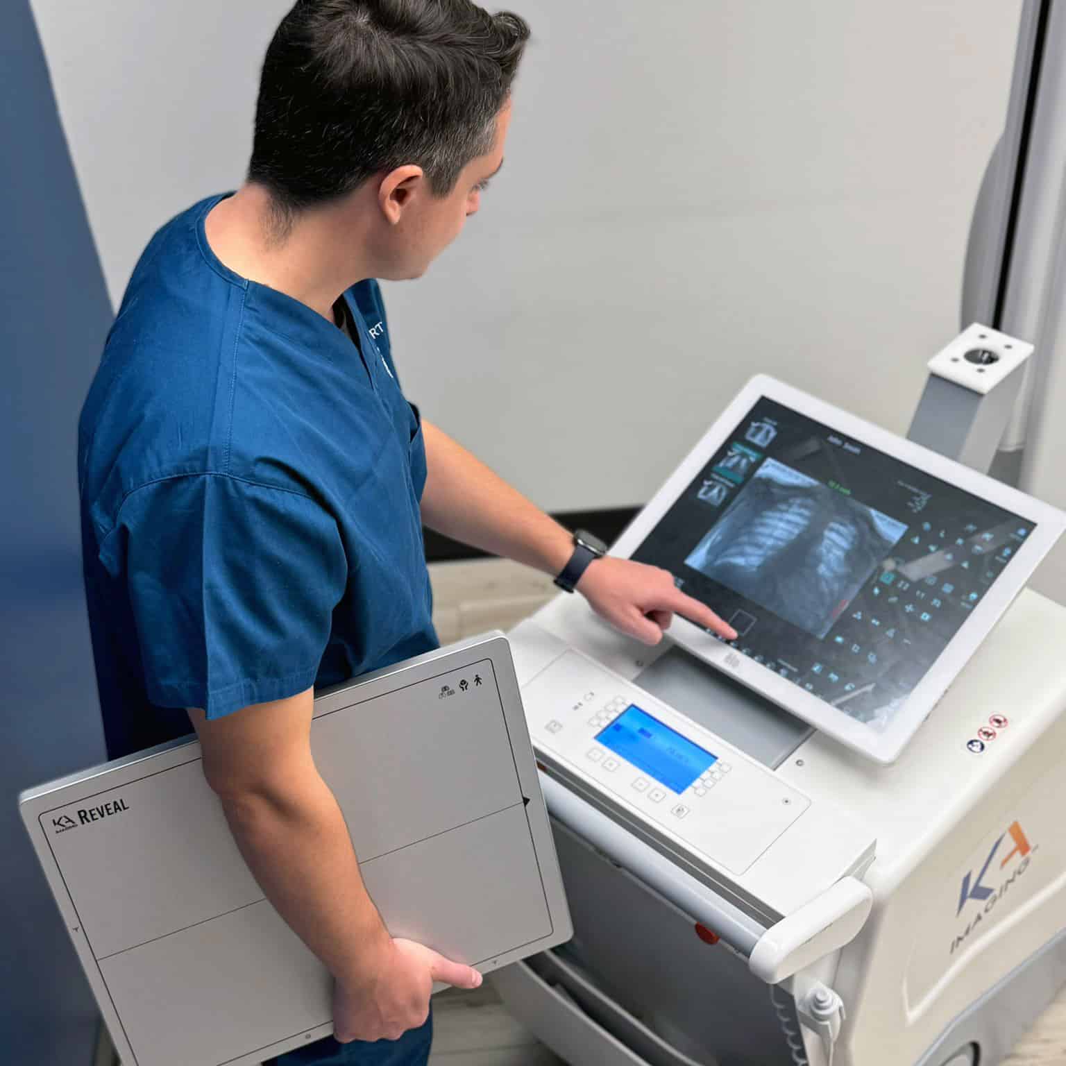
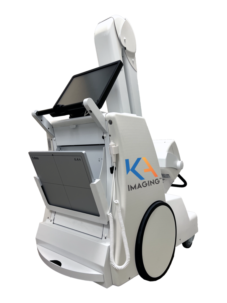
Reveal™ Mobi Pro
The First Premium Mobile X-ray System with Dual-energy Imaging
Reveal™ Mobi Pro is an advanced mobile X-ray system, seamlessly integrating the Reveal™ 35C detector with SpectralDR® dual-energy technology—delivering enhanced diagnostic confidence at the bedside.
By bringing Reveal™ 35C’s SpectralDR® technology to the point of care, Mobi Pro can support more confident diagnoses, which may reduce the need for additional imaging, and potentially lessen patient transports.
Revolutionizing Mobile Radiography
Reveal™ Mobi Pro enhances portable X-ray capabilities by combining high-quality digital radiography with the advanced spectral imaging of the Reveal 35C detector —without requiring additional hardware or extra radiation dose. Designed for critical care environments like the ICU and Emergency Room, it enables accurate diagnosis right at the point of care.
Key Features
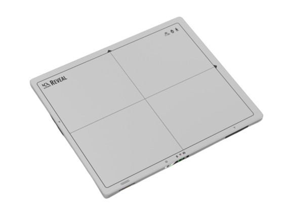
Integrated Reveal™ 35C detector with SpectralDR® technology
Get traditional x-ray, bone and soft tissue images with one exposure
Integrated 19" touch screen monitor with intuitive user interface
Motorized movement, Compact and collapsible column design
8-hour battery life for extended use
Literature
Why choose dual-energy mobile X-ray?
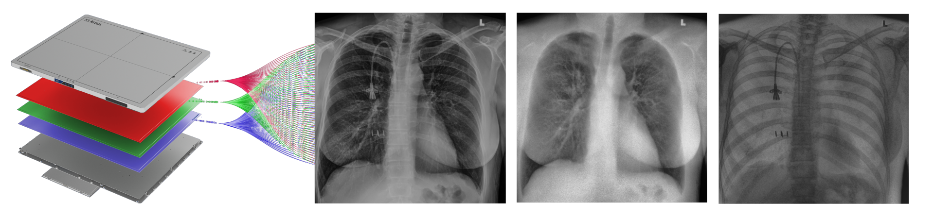
SpectralDR® technology enables simultaneous acquisition of:
Standard X-ray
Soft tissue image
Bone image
—all from a single exposure using a standard X-ray dose.
Dual-energy images are clinically proven1 to:
Improve visibility of lung nodules, pneumonia, pneumothorax, and coronary calcifications
Provide clearer identification of lines, tubes, and retained surgical objects
Improve image clarity without increasing dose or scan time
Impact and ROI
See how integrating dual-energy mobile X-ray could help reduce downstream imaging costs, support earlier, more confident diagnoses, and improve resource utilization in critical care settings.
Clinical Use Cases
Reveal™ Mobi Pro, powered by the Reveal™ 35C detector with SpectralDR® technology, brings the benefits of dual-energy imaging directly to the bedside. From critical care units to emergency departments, the ability to capture traditional, bone, and soft tissue images in a single exposure supports faster, more confident decision-making — without disrupting workflow or increasing radiation dose.
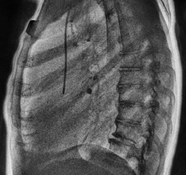
Hidden Pneumonia Detection
A 51-year-old leukemia patient had a normal DR image. The soft tissue image revealed a focal opacity, later confirmed as pneumonia on CT, showing how dual-energy imaging can reveal findings standard X-ray may miss.
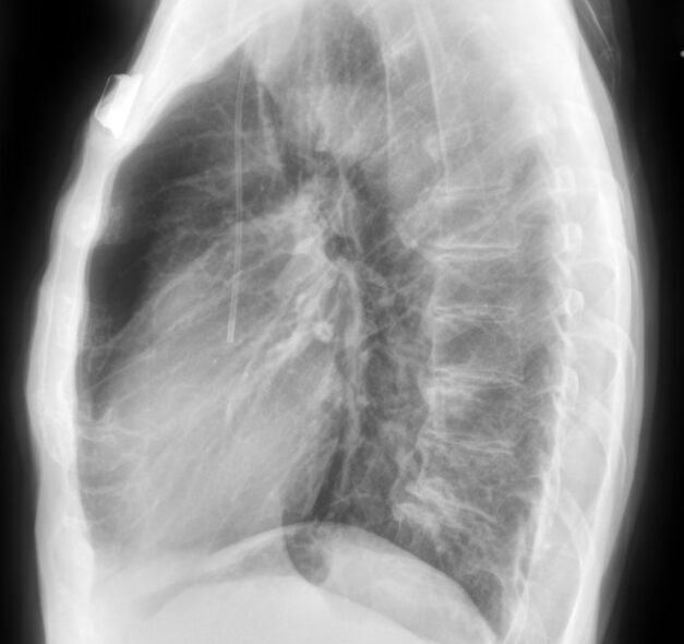
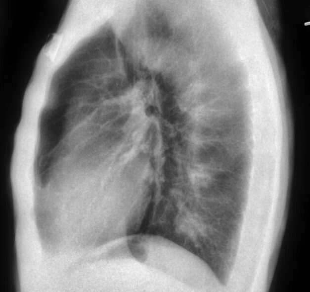
DR/SoftTissue Image


Use the slider left or right to compare DR and soft tissue images.
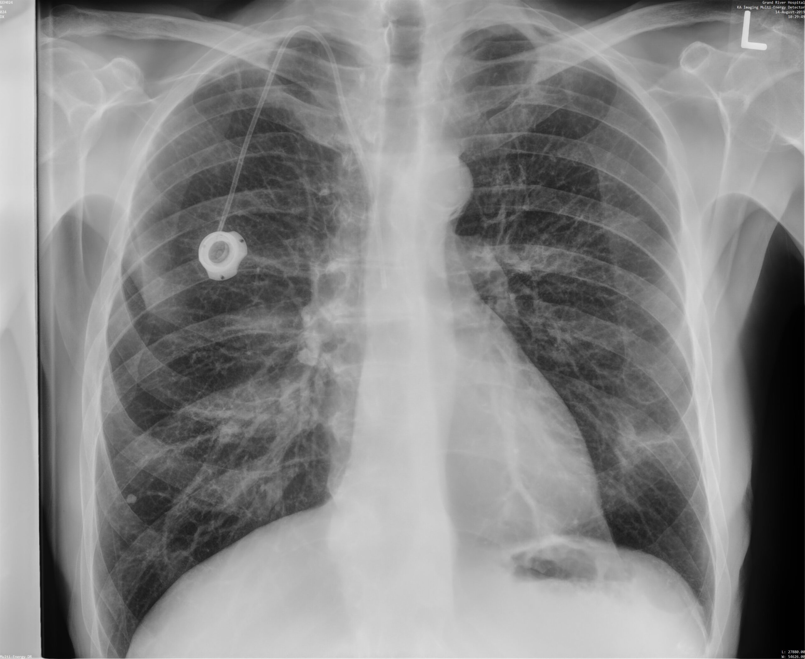
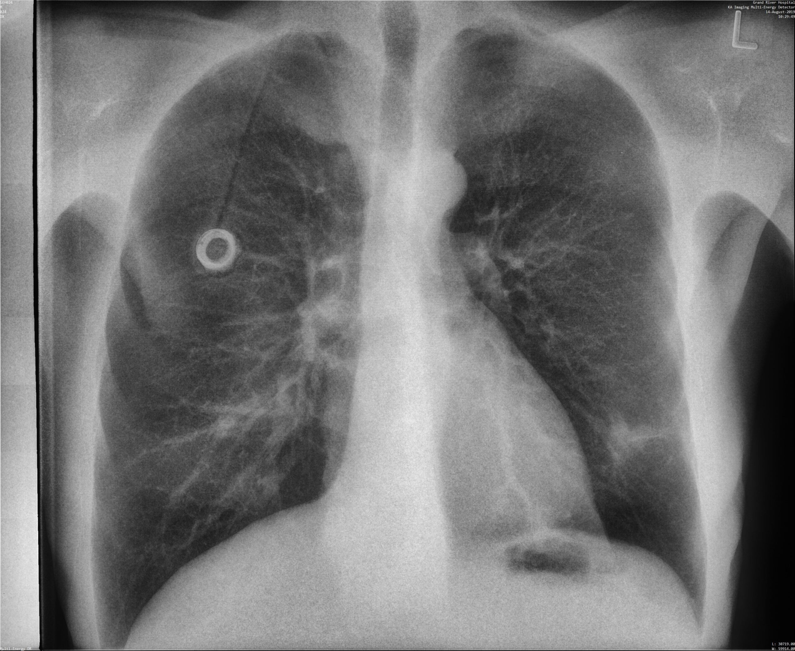
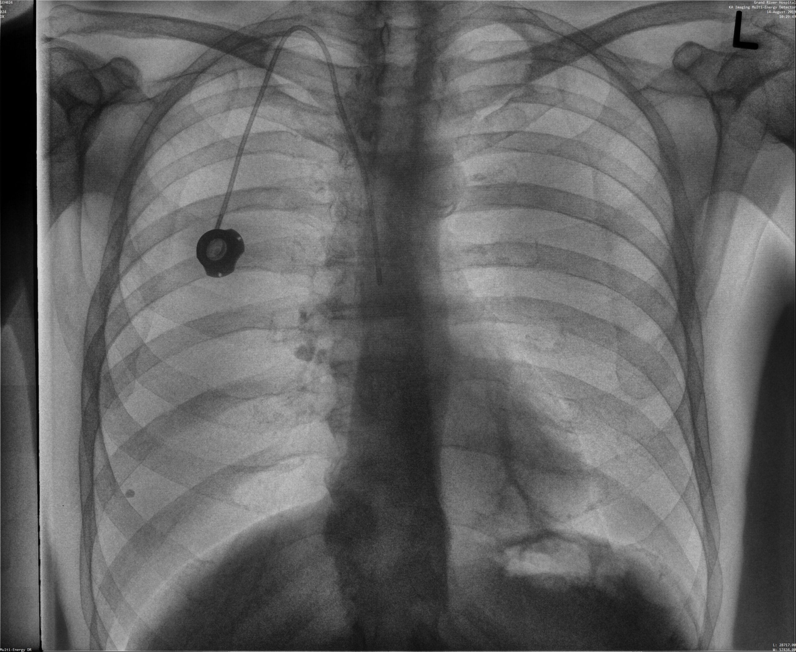
Better Line Placement Confirmation
See vascular wire mesh and tube tips clearly — without additional software, delay, or dose. SpectralDR® streamlines bedside evaluations with confidence and speed.
DR/Bone Image


Use the slider left or right to compare DR and bone images.
Reveal™ 35C: proven results in diagnostic imaging

33% more pneumonia cases found
compared to X-ray thanks to dual-energy images⁴

Increased confidence in 57% of ICU cases
thanks to dual-energy images with no added reading time⁵

43% more lesion visibility
when dual-energy images were included³

Up to 37% decrease
in the number of follow up CT scans for ICU patients9

Up to 61.8% increase in sensitivity
for coronary artery calcium (CAC) compared to standard X-ray, and an AUC of 91% relative to low dose CT10.

Better visibility of lines and tube tips
without extra reading times⁵
Trusted by Users
Media Gallery
Latest blogs
Imaging plays an important role in monitoring patient health and supporting clinical decision-making in intensive…
Tuberculosis remains one of the world’s deadliest infectious diseases. Currently, the critical challenge has shifted…
In 2015, KA Imaging was founded with one major goal: to redefine what X-ray technology…
Reveal Mobi Pro: FDA 510(k), Health Canada, CE Mark.
Contact us for availability in your geographic area.
References:
- Improved patient outcomes for dual-energy subtraction imaging:
1.1 (Lung Nodules) Oda, Seitaro, Kazuo Awai, Yoshinori Funama, Daisuke Utsunomiya, Yumi Yanaga, Koichi Kawanaka, Takeshi Nakaura et al. “Detection of small pulmonary nodules on chest radiographs: efficacy of dual-energy subtraction technique using flat-panel detector chest radiography.” Clinical radiology 65, no. 8 (2010): 609-615.
1.2 (Pneumothorax) Urbaneja, A., Dodin, G., Hoosu, G., et al. (2018) Added Value of Bone Subtraction in Dual-energy Digital Radiography in the Detection of Pneuomothorax: Impact of Reader Expertise and Medical Specialty. The Association of University Radiologists. Elsevier Inc.
1.3 (Pneumonia) Martini, Katharina, Marco Baessler, Stephan Baumueller, and Thomas Frauenfelder. “Diagnostic accuracy and added value of dual-energy subtraction radiography compared to standard conventional radiography using computed tomography as standard of reference.” PloS one 12, no. 3 (2017): e0174285.
1.4 (Tuberculosis) Sharma, Madhurima, Manavjit Singh Sandhu, Ujjwal Gorsi, Dheeraj Gupta, and Niranjan Khandelwal. “Role of digital tomosynthesis and dual energy subtraction digital radiography in detection of parenchymal lesions in active pulmonary tuberculosis.” European Journal of Radiology 84, no. 9 (2015): 1820-1827.
1.5 (Coronary Calcifications) Song, Yingnan, Hao Wu, Di Wen, Bo Zhu, Philipp Graner, Leslie Ciancibello, Haran Rajeswaran et al. “Detection of coronary calcifications with dual energy chest X-rays: clinical evaluation.” The International Journal of Cardiovascular Imaging (2020): 1-8.
- Maurino, S. L., Badano, A., Cunningham, I. A., & Karim, K. S. (2016, March). Theoretical and Monte Carlo optimization of a stacked three-layer flat-panel x-ray imager for applications in multi-spectral diagnostic medical imaging. In Medical Imaging 2016: Physics of Medical Imaging (Vol. 9783, p. 97833Z). International Society for Optics and Photonics.
- S. L. Maurino, K. S. Karim, V. Venkatesh. Diagnostic value of single-exposure dual-energy subtraction radiography in lung lesion detection: initial results. European Congress of Radiology-ECR 2022, 2022.
- Sanchez F, Kandel S, May M, Ronghe S, Rogalla P. Diagnostic value of dual-energy chest x-ray in immunocompromised patients to rule out pneumonia: initial results. European Congress of Radiology-ECR 2021, 2021.
- Rogalla P., Karim K. Added diagnostic value of portable dual-energy chest X-ray in a non-radiological reviewing environment. Radiological Society of North America-RSNA 2022, 2022
- Kuhlman, Janet E., Jannette Collins, Gregory N. Brooks, Donald R. Yandow, and Lynn S. Broderick. “Dual-energy subtraction chest radiography: what to look for beyond calcified nodules.” Radiographics 26, no. 1 (2006): 79-92.
- Manji, Farheen, Jiheng Wang, Geoff Norman, Zhou Wang, and David Koff. “Comparison of dual energy subtraction chest radiography and traditional chest X-rays in the detection of pulmonary nodules.” Quantitative imaging in medicine and surgery 6, no. 1 (2016): 1.
- Karim S Karim, “Single Exposure, Digital Dual-Energy Subtraction X-Ray Ushers in a New Era of Diagnostic X-Ray Imaging,” Radiology Management, Mar/Apr 2021.
- Venkatesh V., Patel N., Clarke K., Karim K. (2024). Implementation experience of a portable single exposure, dual energy X-ray detector for use in the ICU of a community hospital. American Society of Emergency Radiology Meeting.
- Rogalla P, Dos Santos JFP, Wintersperger BJ, et al. Opportunistic Identification of Coronary Artery Calcium and Valve/Vascular Calcifications on Chest X-Ray: Improvements With Single-Exposure Dual-Energy Imaging. Canadian Association of Radiologists Journal. 2024;0(0). doi:10.1177/08465371241291699