
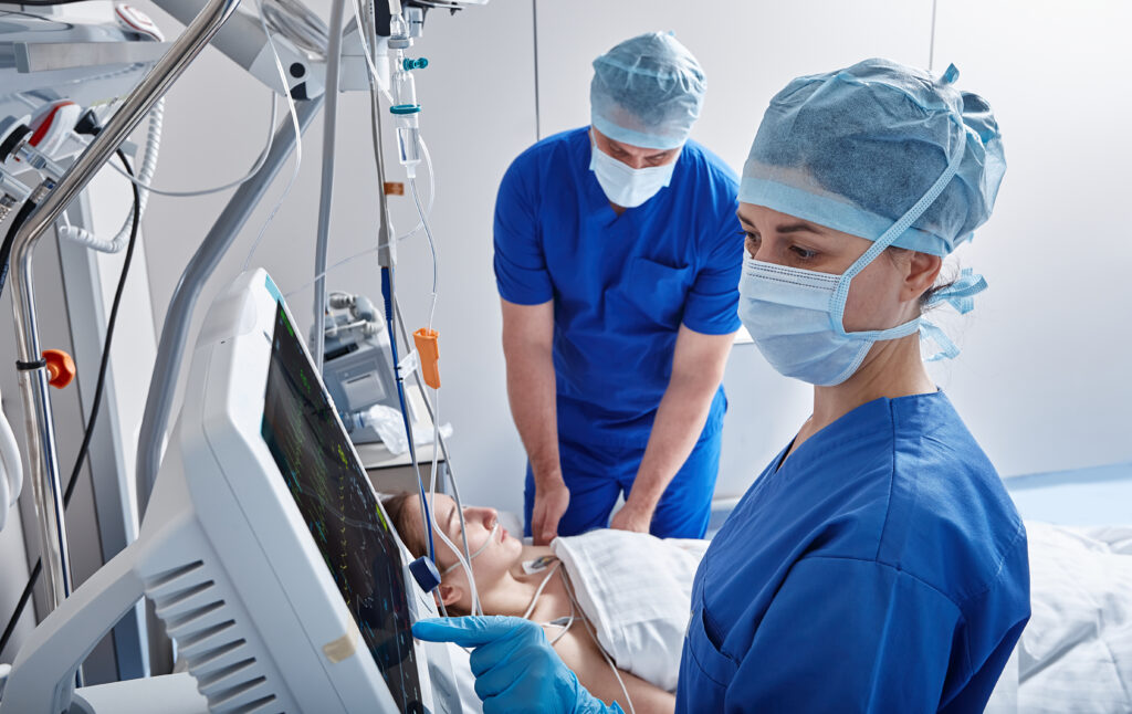
Imaging plays an important role in monitoring patient health and supporting clinical decision-making in intensive care units (ICU). Portable X-ray technologies enable clinicians to provide bedside imaging in the ICU, and these technologies are advancing more and more as time goes on.
For example, spectral imaging is a technique that has the potential to provide additional image information in critical care settings. In portable applications, hospitals can utilize additional spectral information without disrupting clinical workflows.
KA Imaging has developed spectral X-ray imaging technology for portable systems, including the Reveal 35C flat-panel detector powered by SpectralDR® technology. In addition to being portable and retrofittable, the Reveal 35C medical X-ray detector captures dual-energy information with only a single exposure.
This article explores how portable imaging is used in the ICU, why intrahospital transport reduction is an important goal in healthcare, and how spectral X-ray imaging is being studied in bedside imaging environments.
Why Intrahospital Transport Reduction Is an Important Goal in Healthcare
Intrahospital transport is the movement of patients between different areas of the hospital for procedures or imaging exams.
For ICU patients who face specific health risks, these movements require careful coordination between multiple care teams. Intrahospital transport can disrupt workflows, slow down day-to-day operations, and put vulnerable patients at risk.
Because of the risks involved, reducing intrahospital transport is a major goal for healthcare providers in the ICU. The healthcare industry as a whole is continuously evaluating ways to improve workflow efficiency and optimize patient care processes, and intrahospital transport remains one of the biggest challenges in that.
While intrahospital transportation is unavoidable and a necessary part of patient care, reducing transport where it isn’t entirely needed can greatly optimize efficiency and resource utilization.
TLDR; Intrahospital transport reduction is an important goal in healthcare because reducing transport where it isn’t necessary can enable improved workflow efficiency, patient care, and resource utilization.
Benefits of Intrahospital Transport Reduction
- Improved workflow efficiency: Diagnostic evaluations that occur at the bedside can be significantly less disruptive to workflows because it eliminates the need for tight coordination between staff, equipment, and departments.
- Optimized resource utilization: Intrahospital transport typically requires multiple personnel, including nurses, transport staff, and technologists. Bedside imaging removes the need for additional staff on-site to coordinate transport. In the process, hospitals can allocate these resources to higher-priority areas.
- Continuity of care for critically ill patients: Maintaining care within the ICU environment allows clinicians to continue monitoring patients in a familiar environment with specialized equipment and support systems.
It is important to note that there will always be situations where a patient needs to be transported for a more advanced imaging procedure, such as CT. This decision would be made based on clinical judgment and institutional protocols.
The goal with portable ICU imaging is not to completely eliminate intrahospital transport or replace other methods of imaging like CT. Rather, it can help reduce intrahospital transport in scenarios that involve routine imaging procedures.
Intrahospital Transport Risks in ICU Patients
Transporting critically ill patients within the hospital is sometimes necessary for diagnostic procedures or treatments. However, there are many risks involved with transport, including:
- Physiological Instability: ICU patients may require mechanical ventilation, intravenous medications, or hemodynamic support. Movement outside the ICU environment can introduce physiological stress or instability in some patients.
- Monitoring Interruptions: During transport, monitoring equipment may need to be reconfigured or temporarily adjusted. Ensuring continuous patient monitoring throughout transport requires careful coordination.
- Risk of Infection: Moving patients outside the ICU increases exposure to potential hospital-acquired infections (HAIs). The process of transport can expose vulnerable patients to new environments which harbour harmful bacteria, viruses, or other pathogens that could cause infections.
Limiting intrahospital transport wherever possible is an important factor in minimizing these risks and their impact on patient outcomes.
Logistical Challenges in Critical Care Environments
In addition to these risks, there are also logistical challenges that need to be carefully managed. Hospital staff need to coordinate equipment availability, elevator access, hallway clearance, and scheduling within busy radiology departments.
If high-quality bedside imaging is available as an alternative (where applicable), healthcare providers can support clinical workflows while maintaining patient safety.
Clinical Observations from Portable Spectral Chest X-ray in ICU Settings
Spectral radiography is being explored as a complementary tool that may provide additional insight during bedside imaging assessments.
In certain observational settings, the availability of spectral image views has been associated with changes in imaging utilization patterns. For example, some studies have reported up to a 37.5% decrease in chest CT examinations in the ICU in specific clinical scenarios when spectral portable radiography was available.
These findings are observational and context-specific, and may vary depending on clinical workflow and patient population. They suggest that additional image information from spectral radiography may support bedside evaluation and imaging decision-making in certain situations.
It’s important to note that portable spectral X-ray is not intended to replace CT imaging. CT imaging remains a vital part of patient care in the hospital.
TLDR; Clinical observations have found that specific scenarios showed up to 37.5% reduction in chest CTs in the ICU where spectral imaging was available as a suitable alternative. While these studies do suggest that spectral imaging can support hospitals in certain clinical scenarios, it is not meant or encouraged to be used as a replacement for CT.
KA Imaging’s Spectral X-ray: Reveal 35C
KA Imaging has developed spectral X-ray technology designed for integration into portable imaging systems. One example is the Reveal 35C flat-panel detector powered by SpectralDR® technology, which enables the acquisition of conventional and dual-energy images in a single exposure.
This approach produces three image views—standard DR, soft tissue, and bone images—while maintaining workflows and dose levels consistent with standard chest radiography techniques.
Spectral imaging has historically been known for introducing logistical challenges, but Reveal 35C can be integrated into day-to-day operations while maintaining the standard workflow, dose, and techniques of DR imaging. This makes it a potential option for healthcare providers seeking a complementary spectral imaging tool for bedside imaging applications.
You can learn about the capabilities of Reveal 35C and KA’s SpectralDR® technology here: