
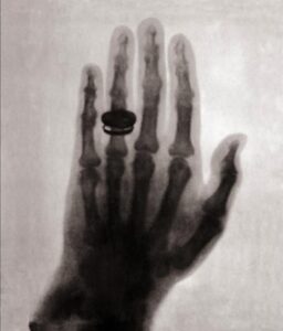
On this day in 1895, Wilhelm Konrad Röntgen discovered the most widely used imaging modality in healthcare today – X-ray. From what started with an accidental discovery and a simple subject of Wilhelm’s wife’s hand, to now the most effective tool in visualizing inner anatomy of subjects for improved diagnoses, X-ray technology has advanced exponentially and elevated the level of healthcare you experience today.
Today’s X-ray (KA Imaging’s patented 𝘚𝘱𝘦𝘤𝘵𝘳𝘢𝘭𝐃𝐑™ technology):
Although in today’s age we draw the connection between X-ray with healthcare, it did not start off this way. Soon after its discovery, people became excited about this new technology and X-ray became a fad that was used in everyday applications – checking shoe sizes, children’s toys, etc. It was only after the second World War that the risks and benefits of the technology were analysed, and we started to use radiation more carefully.
Currently, the ALARA principle guides us in terms of radiation safety. ALARA means “as low as reasonably achievable”. As explained by the Centers of Disease Control and Prevention: “ALARA means avoiding exposure to radiation that does not have a direct benefit to you, even if the dose is small”[1].
Radiography Today
After over one and a quarter centuries of development, we are privileged to have advanced technologies such as that allow radiologists to see more detail in the X-ray images.
KA Imaging’s 𝘚𝘱𝘦𝘤𝘵𝘳𝘢𝘭𝐃𝐑™ technology, featured in Reveal™ 35C, uses the same dose of a traditional DR to capture 3x the image data in the form of 3 spectral images (standard DR, soft tissue, and bone). With the same dose and workflow of a traditional DR system, the supplemental spectral images can aid the physician in visualizing a range of conditions, such as lung nodules, pneumonia, pneumothorax, foreign surgical objects and more[8,9].
Every year on November 8, professionals in the X-ray industry celebrate the discovery of X-ray. Without it, who knows where healthcare would be today. Whether for human or animal healthcare or NDT applications, X-ray allows us to see within the subject for improved analyses and diagnoses. After all, what’s inside matters most.
References
- https://www.cdc.gov/nceh/radiation/alara.html#:~:text=ALARA%20stands%20for%20%E2%80%9Cas%20low,time%2C%20distance%2C%20and%20shielding.
- Britannica – https://www.britannica.com/science/X-ray/Fundamental-characteristics
- FDA – https://www.fda.gov/radiation-emitting-products/medical-imaging/medical-x-ray-imaging
- Government of Canada, Medical X-rays – https://www.canada.ca/en/health-canada/services/health-risks-safety/radiation/medical/x-rays.html
- History of X-rays – https://www.youtube.com/watch?v=fHUzVqoDnts
- NIH – National Institute of Biomedical Imaging and Bioengineering https://www.nibib.nih.gov/science-education/science-topics/x-rays#:~:text=When%20the%20machine%20is%20turned,the%20tissues%20they%20pass%20through.&text=Because%20of%20this%20property%2C%20bones,on%20the%20x%2Dray%20detector
- NHS- https://www.nhs.uk/conditions/x-ray/
- L. Maurino, K. S. Karim, V. Venkatesh. Diagnostic value of single‐exposure dual‐energy subtraction radiography in lung lesion detection: initial results. European Congress of Radiology-ECR 2022, 2022
- Sanchez F, Kandel S, May M, Ronghe S, Rogalla P. Diagnostic value of dual-energy chest x-ray in immunocompromised patients to rule out pneumonia: initial results. European Congress of Radiology-ECR 2021, 2021
- Visit http://kaimaging.com/science-center/reveal-technology/ for more information and references on dual-energy.
