
What is DQE in Radiology?
DQE stands for Detective Quantum Efficiency. Detective Quantum Efficiency measures how efficiently an imaging detector converts X-ray photons into a clear, high-quality image. An X-ray detector’s DQE demonstrates how effectively it uses radiation and preserves the signal-to-noise ratio (SNR).
The formula used to calculate DQE is: DQE = (SNR_out)^2 / (SNR_in)^2.
Why Does it Matter?
DQE matters because it measures two major aspects of an X-ray image:
- Image quality, which can affect whether the image is viable for diagnostic purposes. High DQE images are clear, sharp, and able to visualize finer details like subtle changes in soft tissue.
- The radiation dose needed for a high-quality image. High DQE imaging systems can generate higher quality images with less radiation than a lower DQE system. Radiation dosages are an important consideration because too much radiation can negatively impact patient safety.
High DQE vs. Low DQE: What is the Difference?
High DQE vs. Low DQE
- Image Quality
- High DQE: Generates clear and detailed images
- Low DQE: Generates images with more noise and less detail
- Radiation Dose
- High DQE: Requires less radiation to get an image suitable for diagnostics
- Low DQE: Higher radiation dosage needed to be viable for diagnostics
- Efficiency
- High DQE: Converts more useful X-ray photons
- Low DQE: Converts less X-ray photons, resulting in lower quality
- Patient Safety
- High DQE: Safer for patients because it requires less radiation
- Low DQE: Not as safe because patients are exposed to more radiation
- Repeat Imaging
- High DQE: More reliable and generally requires less repeat imaging
- Low DQE: May require repeat imaging due to poor image quality
- Suitability for Vulnerable Patients
- High DQE: Works better for pediatric patients or those more sensitive to radiation
- Low DQE: Less suitable for pediatrics or radiation-sensitive patients
Benefits of a High DQE X-ray Detector
A high DQE X-ray detector is the optimal choice for diagnostic imaging purposes. But why? Here are a few of the major benefits that come with using a high DQE X-ray detector.
Higher Image Quality & Signal-to-Noise Ratio
When using X-ray for diagnostic purposes, higher image quality will always be better. High-quality X-ray images with high-contrast allow radiologists to more easily detect subtle abnormalities, such as lung nodules. This makes it less likely that a radiologist will misinterpret an image and give inaccurate results.
High DQE X-ray has potential to enable faster and more accurate diagnosis. Clearer images make results less ambiguous, meaning reading time could be reduced. This is valuable for emergency settings.
Moreover, these images have a higher signal-to-noise ratio (SNR). For SNR, the signal represents what you are trying to capture, while noise is random and interferes with the desired signal.. Noise can obscure critical diagnostic information, which is why a higher SNR is ideal in practically every medical scenario.
Less Radiation
Since high DQE detectors do a better job of converting X-ray photons into usable image signals, less radiation is needed for diagnostic-quality imaging. A few of the ways this benefits day-to-day medical operations is:
- Reducing repeat imaging, which is sometimes done to capture a better diagnostic image. High DQE images can eliminate the need for retakes, thereby reducing the overall radiation exposure.
- Patients with chronic conditions may require ongoing X-ray imaging. When repeat imaging is needed, there is less radiation risk with high DQE imaging than low DQE imaging.
- Children and pregnant patients are more vulnerable to ionizing radiation. The risks are reduced by using less radiation for a high DQE X-ray.
Better for Mobile and Portable Imaging
High DQE portable imaging is crucial for ICU, emergency, field hospital, and disaster relief settings where patient movement is limited and low-dose imaging is essential. Often, X-ray imaging will be needed in non-ideal conditions. High DQE systems are the best choice for these scenarios, as they can quickly capture high-quality diagnostic images with the standard radiation dose.
Furthermore, retakes are not always possible in these settings, which is why it’s crucial to get a diagnostic-quality image the first time.
Cost-Effective
High DQE imaging can lead to better savings on day-to-day operational costs and reduce resource spending. The efficiency offered by high DQE X-ray detectors is able to speed up the imaging process with fewer technical adjustments and retakes needed.
Similarly, reading time is often reduced because high DQE images demonstrate higher image quality that enhances low contrast areas and captures fine details.
What is an X-ray detector with a high DQE?
The Reveal™ 35C flat panel detector, developed in Canada by KA Imaging, sets the benchmark for high DQE performance in mobile and portable X-ray imaging. Featuring a DQE of up to 75% at 0 lp/mm, Reveal 35C is designed to deliver superior image quality while minimizing radiation exposure. This makes it suitable for ICU, emergency, and other environments. It is the first and only mobile dual-energy detector on the market.
High DQE Imaging with Reveal 35C
Reveal 35C has a triple layer design which simultaneously captures three images in a single exposure, all without motion artifacts. To do this, the Reveal 35C only requires the standard radiation dose. The dosage does not need to be increased to achieve dual-energy imaging.
Of the three images is a traditional DR image which captures both bone and soft tissue, as well as supplemental bone and soft tissue images. This enables easy material differentiation for radiologists.
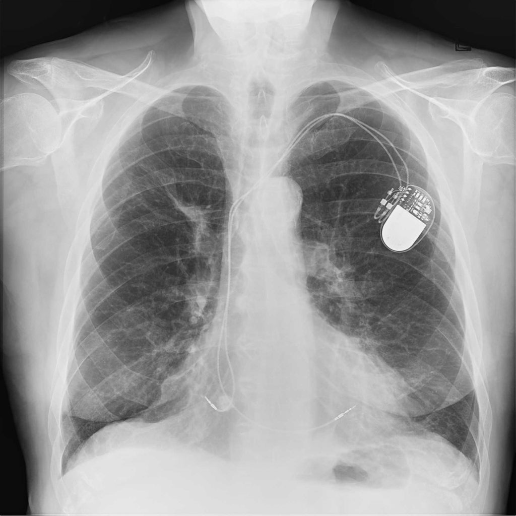
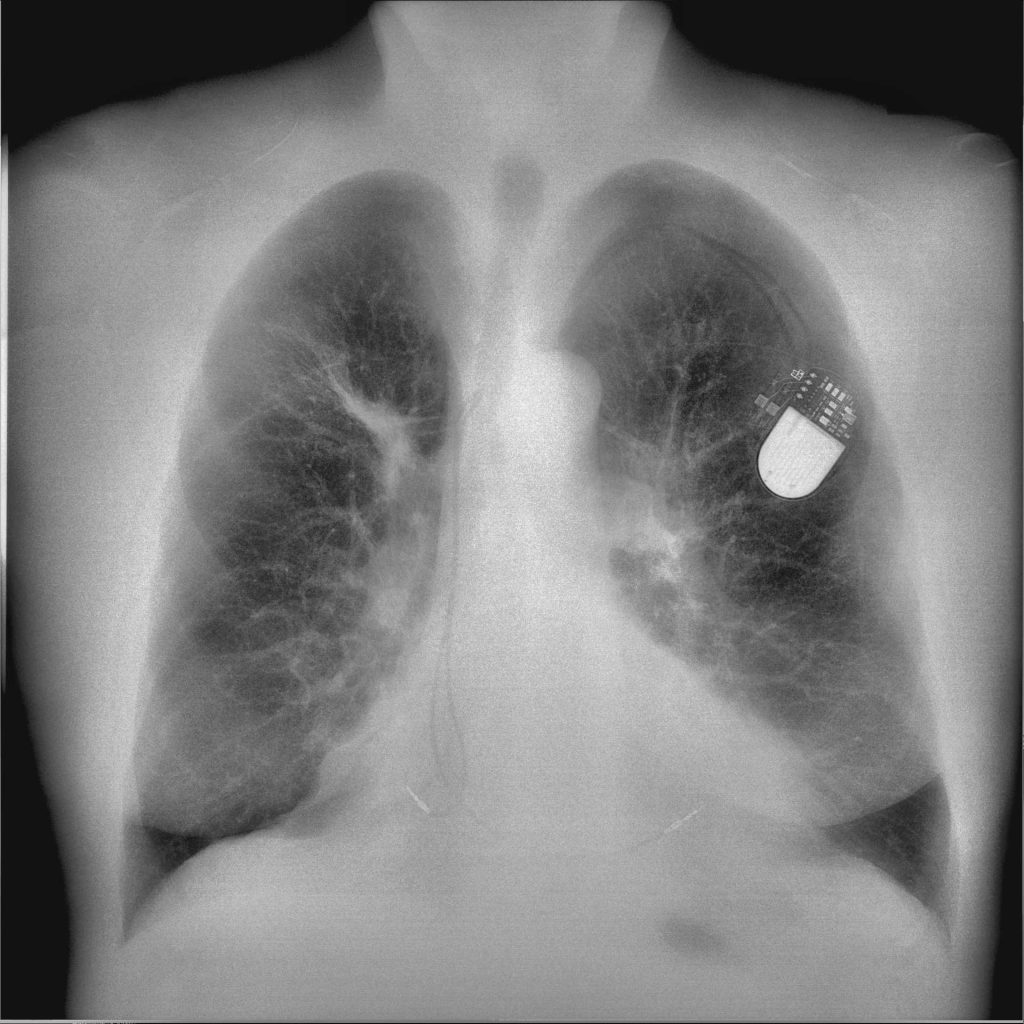
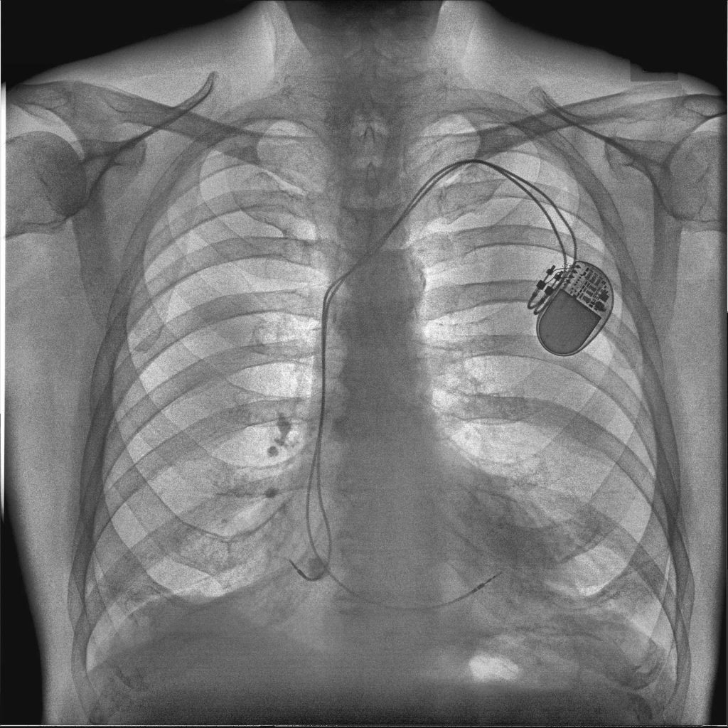
On the left is the traditional DR image, in the middle is the soft tissue image, and on the right is the bone image.
What is Reveal 35C’s DQE?
The Reveal 35C flat panel detector has up to 75% DQE, among the highest in its class.
Conclusion: Why DQE Matters More Than Ever
As diagnostic imaging continues to evolve, understanding metrics like Detective Quantum Efficiency is essential for making informed decisions about equipment, safety, and clinical performance. High DQE detectors like Reveal™ 35C not only deliver sharper, more reliable images – they also support safer imaging practices with lower radiation exposure and fewer retakes. In environments where speed, accuracy, and patient safety are paramount, a detector with high DQE performance – like Reveal™ 35C – offers clear advantages that support efficient and confident decision-making.
Frequently Asked Questions about Detective Quantum Efficiency
What is the difference between DQE and MTF?
Both DQE (Detective Quantum Efficiency) and MTF (Modulation Transfer Function) are methods for measuring image quality, but the way they are measured is different.
DQE measures how efficiently an imaging system converts X-ray photons into a useful image.
MTF measures an imaging system’s ability to depict contrast at different spatial resolutions.
For example, an imaging system with poor MTF may clearly show large structures, but have trouble maintaining that same contrast with the finer details of that image.
What increases DQE?
Some of the ways you can increase DQE in an imaging system are by:
- Using highly-sensitive detector materials (ex. cesium iodide)
- Optimizing pixel design for better signal capture
- Using advanced electronics to reduce electronic noise
- Using structured scintillators which help reduce light scatter
All of these practices help imaging systems more efficiently convert X-ray photons into a high-quality image.
Does a higher DQE always mean better image quality?
Not necessarily. While DQE reflects a system’s efficiency in converting X-rays into signal, image quality also depends on factors like MTF, scatter, and clinical application. DQE should be considered alongside other performance indicators.
How is DQE measured, and at what frequencies?
DQE is typically measured at different spatial frequencies (e.g., 0, 1, 2 line pairs/mm). A full DQE curve provides a more accurate picture of performance across fine and coarse detail.
Is there a tradeoff between DQE and resolution?
Sometimes. Detectors with smaller pixels offer higher resolution but can have lower DQE due to increased noise. Advanced designs, like those in Reveal 35C, optimize both DQE and resolution.
What clinical scenarios benefit most from high DQE?
High DQE is particularly valuable in portable/mobile X-ray, ICU, pediatric, thoracic, and emergency imaging—anywhere diagnostic quality is needed under dose or time constraints.
What’s the difference between DQE and CNR (Contrast-to-Noise Ratio)?
DQE measures efficiency of signal conversion; CNR assesses how well contrast differences stand out from noise in a specific image. Both impact diagnostic clarity but reflect different properties.
How does the Reveal 35C’s DQE compare to other detectors?
Reveal 35C offers up to 75% DQE, among the highest in its class. Its high DQE, combined with dual-energy capability, supports superior diagnostic utility at lower radiation doses.