
BrillianSe™ X-ray Detector

BrillianSe™ is our patented amorphous selenium (a-Se) X-ray detector for high-brilliance imaging, expanding the horizons of scientific imaging and non-destructive testing (NDT). BrillianSe™ provides a unique combination of high spatial resolution using 8 μm pixels, and high Detective Quantum Efficiency (DQE) for energies up to 110 keV. This combination enables efficient imaging at low flux and high energy, as well as propagation-based (grating less) in-line phase-contrast enhancement for improved sensitivity when imaging low-density materials.
Product overview
Sensor Type: amorphous Selenium with CMOS APS
8 μm Pixel Pitch
~36% at 10 cycles/mm at 60 KV (2 mm al filtration) DQE
Propagation-based X-ray phase contrast
Literature
BrillianSe™: Taking Scientific Imaging and Non-Destructive Testing (NDT) to the Next Level
BrillianSe™, our innovative amorphous selenium (a-Se) X-ray detector, revolutionizes high-brilliance imaging by delivering unparalleled clarity and precision. This technology opens new frontiers in scientific research and non-destructive testing (NDT) for various applications:
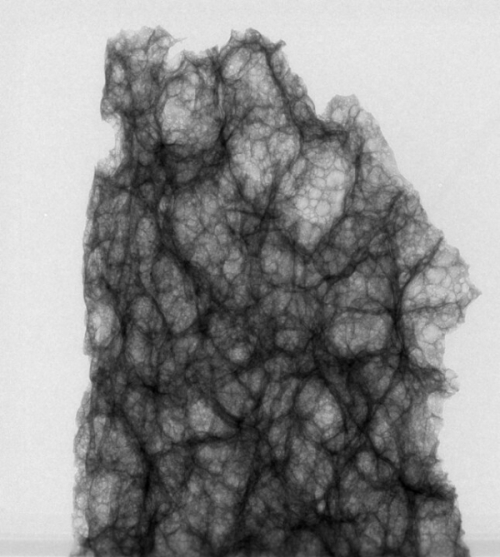
Low-density material phase-contrast
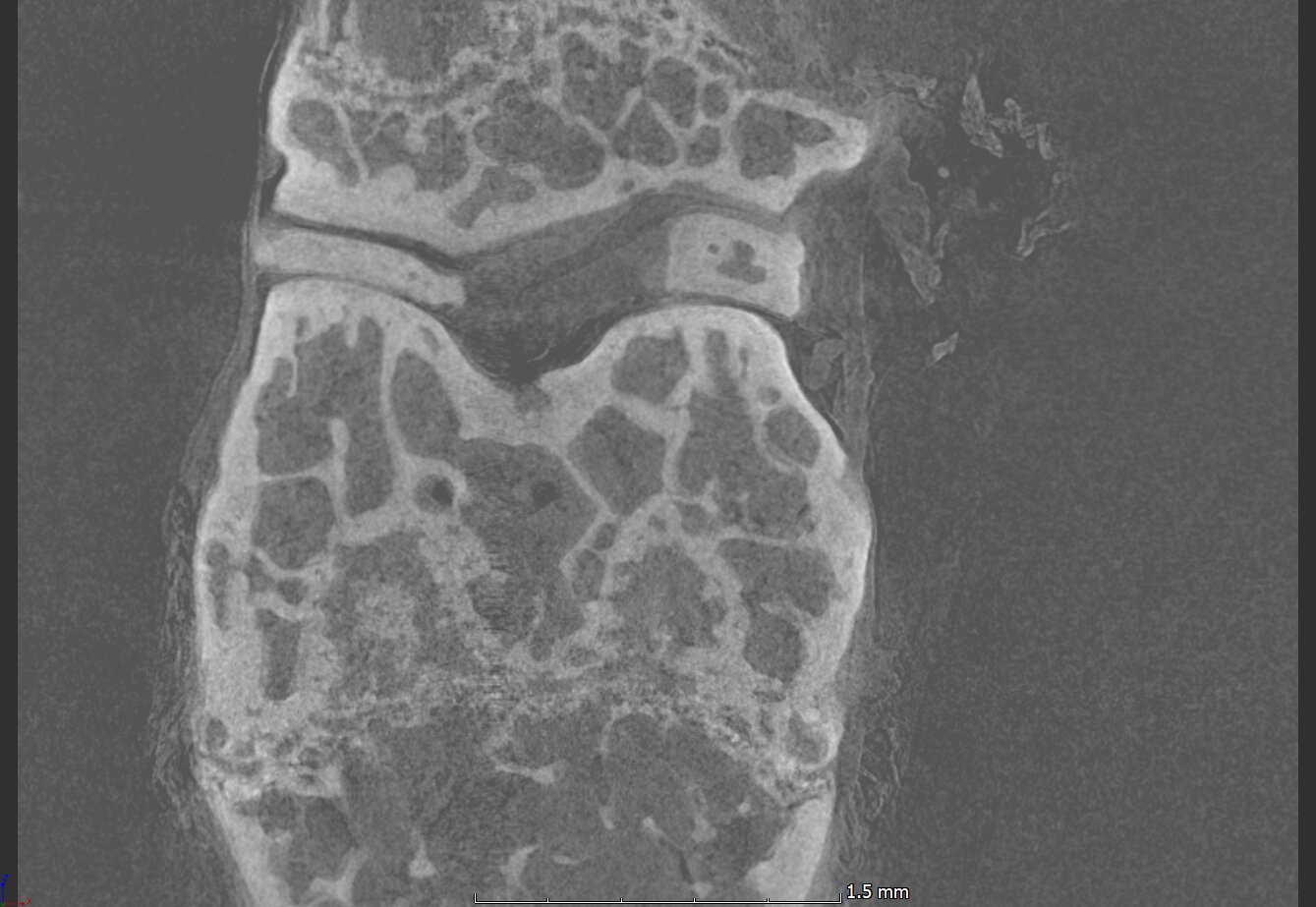
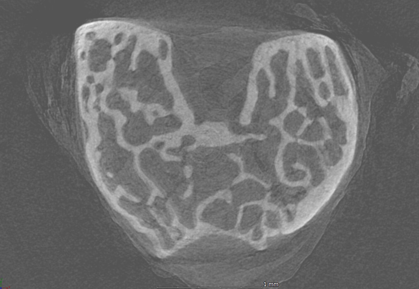
Synchrotron micro-CT
Single photon sensitivity (>50 keV)
High energy (>50 keV) Bragg Coherent Diffraction Imaging
Customer Stories
How BrillianSe™ works
The hybrid a-Se/CMOS detector uses an a-Se photoconductor with high intrinsic spatial resolution for direct conversion of X-ray photons to electric charge. The electronic signal is then read out by a low noise CMOS active pixel sensor (APS). Without the need to first convert X-ray photons to visible light, as in indirect scintillator-based approaches, thinning of the conversion layer to minimize optical scatter is not necessary.


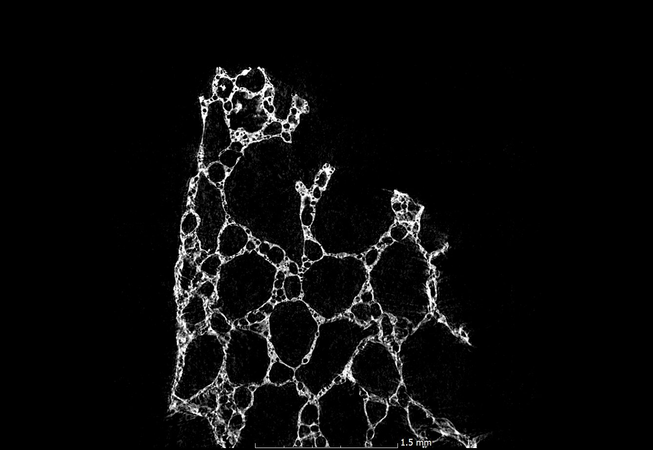
Phase Contrast: better visualization for low-density materials
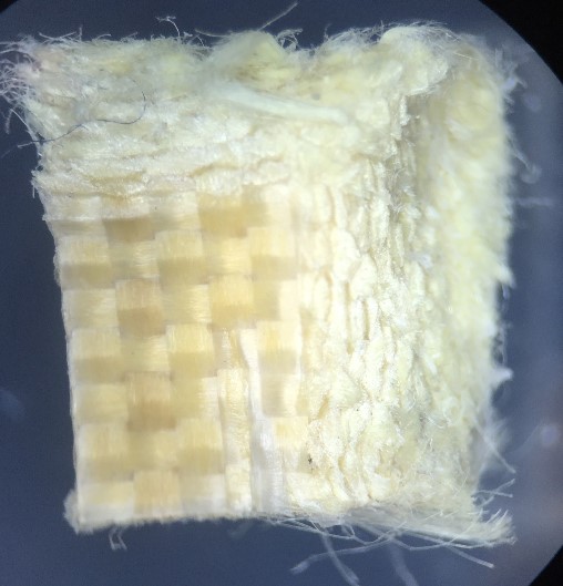
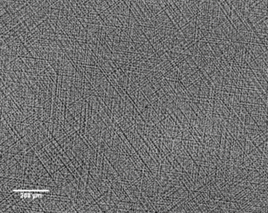
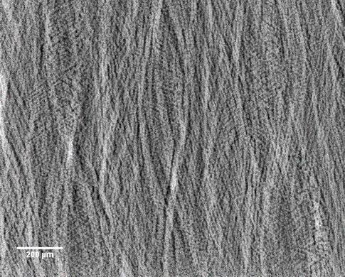
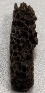
BrillianSe™ excels in phase-contrast imaging, which is crucial when traditional X-ray methods struggle with low-density materials like soft tissues or polymers. Unlike conventional detectors that measure only X-ray intensity, BrillianSe™ can capture subtle phase shifts, enhancing image contrast. This is achieved using free-space propagation, where phase variations translate directly into intensity fluctuations, offering clearer, more detailed images.
Latest blogs
KA Imaging’s patented amorphous selenium (a-Se) BrillianSe™ X-ray imaging detectors have been designed into the…
In the realm of industrial safety and quality assurance, Non-destructive Testing (NDT) emerges as a…
Original research
S. Abbaszadeh, CC Scott, O Bubon, A Reznik, KS Karim, “Enhanced detection efficiency of direct conversion X-ray detector using polyimide as hole-blocking layer,” Scientific Reports, December 2013
G.P. Lindberg, T. O’Loughlin, N. Gross, A. Mishchenko, A. Reznik, S. Abbaszadeh, K.S. Karim, G. Belev, B. Weinstein, “Photo-crystallization in a-Se layer structures: effects of film-substrate interface-rigidity,” Journal of Applied Physics, vol. 116, November 2014.
A. Parsafar, C. Scott, A. El-Falou, P. Levine, K.S. Karim, “Direct-Conversion CMOS X-Ray Imager with 5.6 μm × 6.25 μm Pixels,” IEEE Electron Device Letters, 36(5), pp. 481-3, May 2015.
C.C. Scott, A. Parsafar, A. El-Falou, P. M. Levine, K.S. Karim, “High Dose Efficiency, Ultra-high Resolution Amorphous Selenium/CMOS Hybrid Digital X-ray Imager,” IEEE International Electron Devices Meeting (IEDM) Technical Digest, December 2015.
Related research
- Comparing conventional and phase contrast imaging
Gureyev, Timur Eugenievich, Sheridan C. Mayo, Damian E. Myers, Ya Nesterets, D. M. Paganin, A. Pogany, Andrew W. Stevenson, and S. W. Wilkins. “Refracting Röntgen’s rays: propagation-based x-ray phase contrast for biomedical imaging.” Journal of Applied Physics 105, no. 10 (2009): 102005.
Krenkel, Martin, Mareike Töpperwien, Christian Dullin, Frauke Alves, and Tim Salditt. “Propagation-based phase-contrast tomography for high-resolution lung imaging with laboratory sources.” AIP Advances 6, no. 3 (2016): 035007.
Olivo, A., and E. Castelli. “X-ray phase contrast imaging: From synchrotrons to conventional sources.” La Rivista Del Nuovo Cimento 37 (2014): 467-508.
Bravin, Alberto, Paola Coan, and Pekka Suortti. “X-ray phase-contrast imaging: from pre-clinical applications towards clinics.” Physics in Medicine & Biology 58, no. 1 (2012): R1.
Bidola, P., K. Morgan, M. Willner, A. Fehringer, S. Allner, F. Prade, F. Pfeiffer, and K. Achterhold. “Application of sensitive, high‐resolution imaging at a commercial lab‐based X‐ray micro‐CT system using propagation‐based phase retrieval.” Journal of Microscopy 266, no. 2 (2017): 211-220.
- Propagation
Wilkins, S. W., T. Ei Gureyev, D. Gao, A. Pogany, and A. W. Stevenson. “Phase-contrast imaging using polychromatic hard X-rays.” Nature 384, no. 6607 (1996): 335-338.
Mayo, Sheridan C., Andrew W. Stevenson, and Stephen W. Wilkins. “In-line phase-contrast X-ray imaging and tomography for materials science.” Materials 5, no. 5 (2012): 937-965.
- Gratings
Pfeiffer, Franz, Timm Weitkamp, Oliver Bunk, and Christian David. “Phase retrieval and differential phase-contrast imaging with low-brilliance X-ray sources.” Nature physics 2, no. 4 (2006): 258-261.