Entries by admin

, ,

KA Imaging Signs Australian Distributor – Regional Health Care Group

Launching KA Imaging into a $4B AUD market Waterloo, Ont. (June 08, 2021) Medical distribution company Regional Health Care Group (RHCG) will now be bringing KA Imaging’s innovative X-ray technologies to Australia and New Zealand. This agreement ensures sole rights to RHCG as KA Imaging’s distributor. “This is a fantastic opportunity for KA Imaging that moves us towards fulfilling our vision of innovative […]

, ,

Bio Scan Signed as KA Imaging’s Sole Mexican Distributor

Bringing Innovative X-ray Solutions to a Market Forecast to Reach $340.2 Million Next Year  Waterloo, Ont. (May 31, 2021) – KA Imaging has announced a distribution agreement with Bio Scan S.A. de C.V. in Mexico. Bio Scan, as the sole distributor in Mexico, will be responsible for sales and support in full collaboration with KA Imaging territory-wide. […]

, ,

KA Imaging signs with Alpha Imaging for distribution in the USA

Alpha Imaging is one of the largest in the country serving 14 states in the Northeast, Mid-Atlantic and Central US 


WATERLOO. ON (May 25, 2021)- KA Imaging has signed a distribution agreement in the USA with Alpha Imaging. Hospitals, medical facilities, and practices from 14 states can now acquire Reveal 35C through Alpha Imaging, one of the largest distributors of medical imaging equipment in the country. 

Robert Moccia, VP of Sales for KA Imaging in the United States and Canada, is thrilled to have made this agreement.  

“Our partnership with Alpha is a big step towards KA Imaging’s vision to bring innovative X-ray everywhere. Alpha Imaging’s 30+ year track record of providing Imaging equipment to some of the most prestigious healthcare institutions in the US gives KA Imaging the access and credibility to excite the market with our technology and ultimately improve patient care” said Moccia.  

Michael Perrico, CEO of Alpha Imaging, said: “The addition of KA Imaging’s Reveal 35C adds yet another advanced imaging technology to our robust portfolio of medical imaging offerings. Single exposure subtracted chest imaging will provide clients with a low dose and cost-effective alternative to traditional dual energy systems. We are excited to partner with KA Imaging.” 

Reveal 35C is the world’s first and only portable dual-energy X-ray detector. The detector provides radiologists with three unobstructed images in a single shot that are free from motion artifacts. Reveal is currently being used in two clinical trials, for detecting both lung cancer and pneumonia (including COVID-19) with great initial success. 

About Alpha Imaging:
Alpha Imaging LLC, headquartered in Ohio, is one of the largest independent sales and service providers of advanced medical imaging equipment in the United States. Alpha is celebrating over 30 years in the imaging industry and Alpha Imaging partners with leading global manufacturers to deliver state-of-the-art products and services that meet the clinical, operational and economic needs of the U.S. healthcare market. One of Inc. Magazine’s fastest growing companies for 10 straight years, Alpha Imaging builds customer loyalty with a personal touch rarely seen in today’s medical imaging marketplace. For more information, visit https://alpha-imaging.com/ 


About KA Imaging:
A spin-off from the University of Waterloo, KA Imaging specializes in developing innovative X-ray imaging technologies and systems, providing solutions to the medical, veterinary, and non-destructive test industrial markets. For more information, visit www.kaimaging.com. 


Contact: 

Fernanda Fraga  

Media Relations 
ffraga@kaimaging.com  
T: 226.215.9897 

,

Reveal™ 35C Clinical Case Series: (Pneumonia)

Welcome to the new Clinical Case Series

In KA Imaging’s new Clinical Case Series, the focus will be on diagnosing pneumonia, considering the growing need for efficient diagnosis in COVID-19 patients. KA Imaging’s Reveal™ 35C is currently being used in two clinical trials in the University Health Network (UHN).

1. Focal Opacity in RUL

In this video, Dr. Karim S. Karim demonstrates the differences between traditional X-ray, soft tissue, and CT images. The traditional chest X-ray image in this case was deemed normal by a radiologist, but the dual-energy subtraction soft tissue image taken by Reveal™ 35C showed a focal opacity in the right lobe indicating pneumonia.

2. Left Perihilar Opacity

In this case, learn how Reveal™ 35C helped a radiologist enable visuals of perihilar opacities in order to diagnose fungal pneumonia with confidence.

,

KA Imaging Installs Two New Mobile X-ray Units At University Health Network

New units support early detection of COVID-19 pneumonia 

WATERLOO, ON (May 17, 2021) 

KA Imaging, a developer of innovative X-ray imaging technologies and systems, today announced that it has installed two new mobile X-ray units within the University Health Network (UHN) to enhance current clinical studies and support patient care. One new unit is being used at the Princess Margaret Cancer Centre, and the other at the Toronto General Hospital.  

Clinical testing began in September, 2020, at Princess Margaret Cancer Centre with two novel technologies, one being KA Imaging’s Reveal™, a portable, dual-energy X-ray solution, in a study pertaining to the early detection of pneumonia including COVID-19. 

 “In the first patients, Reveal’s dual-energy capabilities have already helped find pneumonia with higher confidence. We wanted to add another system to increase our imaging capacity,” said Patrik Rogalla, MD, site director, Princess Margaret Hospital, Department of Medical Imaging, and the study’s principal investigator.  

Reveal provides radiologists with unobstructed views of the lungs to aid in the visualization of pneumonia, and many other clinical indications, such as fractures, cardiac calcifications or masses. 

 “This technology provides three images with a single shot that are all free from motion artifacts,” said Dr. Karim S. Karim, CTO of KA Imaging. “And Reveal is uniquely portable and mobile, so it can be taken to a patient’s bedside, further minimizing the risk of viral spread.” 

“The fact that it is a mobile system will make a difference for us, as we will be able to get the X-ray to the patients, which is particularly helpful in a COVID-19 scenario and also for patients with mobility issues,” said Dr. Rogalla. 

“Our partnership with UHN gives true meaning to years of dedicated work. To see such promising initial results from Reveal means that our goal to positively impact healthcare is being achieved,” said KA Imaging President and CEO Amol Karnick. “We want to provide innovative X-ray solutions and change lives, and we are confident that Reveal can help in the early detection of COVID-19 pneumonia.”  

About KA Imaging 

A spin-off from the University of Waterloo, KA Imaging specializes in developing innovative X-ray imaging technologies and systems, providing solutions to the medical, veterinary, and non-destructive test industrial markets. For more information, please visit www.kaimaging.com. 

Contact: 

Fernanda Fraga  

Media Relations
ffraga@kaimaging.com 
T: 226.215.9897 


,

How KA Imaging’s Dual-energy Solution Sets a New Industry Standard

Reveal™ 35C is available in Canada and the USA. Contact us for inquiries and availability in other countries.

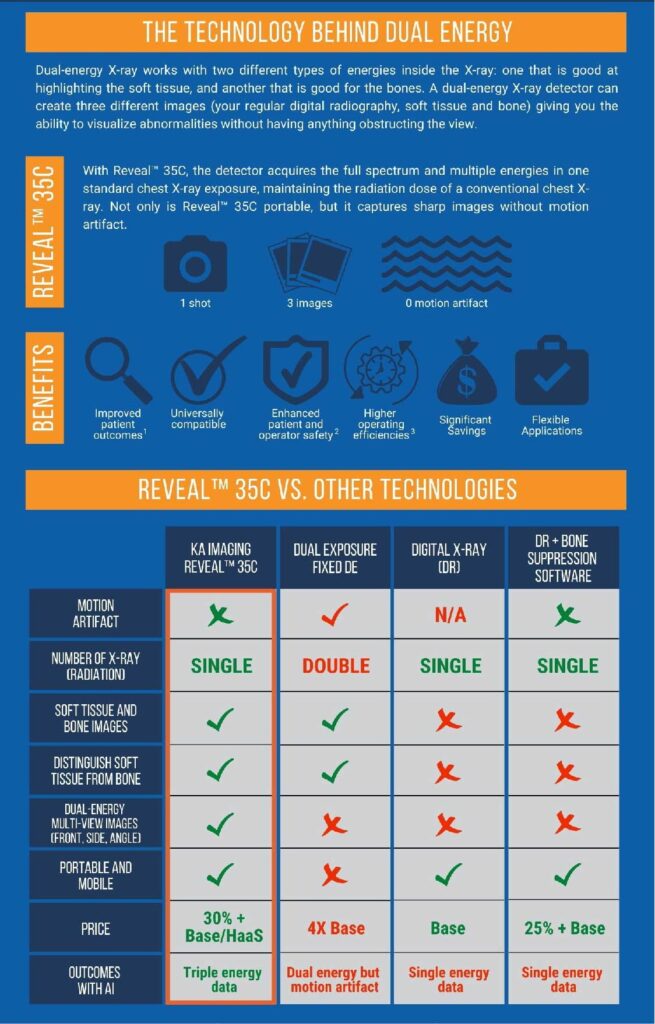
Reveal™ 35C is the world’s first and only single exposure, portable, digital dual-energy subtraction (DES) X-ray detector that overcomes previous DES technical limitations. This detector raised the possibility of a DES retrofit in existing digital X-ray rooms. The portability of a single exposure DES detector also allows for point-of-care high-quality diagnostic imaging to increase access in underserved regions and low mobility patients.

Dual-energy has been proven to detect more solitary pulmonary nodules, pneumothorax, pneumonia, tuberculosis, and coronary calcium than conventional analog or digital chest X-rays. This early detection enables faster/improved patient outcomes, additional hospital revenue, cost savings to healthcare systems, and reduces exposure to malpractice claims. Additionally, the Reveal™ 35C has 20X less radiation compared to CT scans and reduces radiologist reading time for X-rays by 30% at 10X lower purchase and operating costs than traditional CT scans.


VISIT THE PRODUCT PAGE

References


1. Improved patient outcomes


(Lung Nodules) Oda, Seitaro, Kazuo Awai, Yoshinori Funama, Daisuke Utsunomiya, Yumi Yanaga, Koichi Kawanaka, Takeshi Nakaura et al. “Detection of small pulmonary nodules on chest radiographs: efficacy of dual-energy subtraction technique using flat-panel detector chest radiography.” Clinical radiology 65, no. 8 (2010): 609-615.


(Pneumothorax) Urbaneja, A., Dodin, G., Hoosu, G., et al. (2018) Added Value of Bone Subtraction in Dual-energy Digital Radiography in the Detection of Pneuomothorax: Impact of Reader Expertise and Medical Specialty. The Association of University Radiologists. Elsevier Inc.


(Pneumonia) Martini, Katharina, Marco Baessler, Stephan Baumueller, and Thomas Frauenfelder. “Diagnostic accuracy and added value of dual-energy subtraction radiography compared to standard conventional radiography using computed tomography as standard of reference.” PloS one 12, no. 3 (2017): e0174285.


(Tuberculosis) Sharma, Madhurima, Manavjit Singh Sandhu, Ujjwal Gorsi, Dheeraj Gupta, and Niranjan Khandelwal. “Role of digital tomosynthesis and dual energy subtraction digital radiography in detection of parenchymal lesions in active pulmonary tuberculosis.” European Journal of Radiology 84, no. 9 (2015): 1820-1827.


(Coronary Calcifications) Song, Yingnan, Hao Wu, Di Wen, Bo Zhu, Philipp Graner, Leslie Ciancibello, Haran Rajeswaran et al. “Detection of coronary calcifications with dual energy chest X-rays: clinical evaluation.” The International Journal of Cardiovascular Imaging (2020): 1-8.


Kuhlman, Janet E., Jannette Collins, Gregory N. Brooks, Donald R. Yandow, and Lynn S. Broderick. “Dual-energy subtraction chest radiography: what to look for beyond calcified nodules.” Radiographics 26, no. 1 (2006): 79-92.


2. Enhanced patient and operator safety.


Kuhlman, Janet E., Jannette Collins, Gregory N. Brooks, Donald R. Yandow, and Lynn S. Broderick. “Dual-energy subtraction chest radiography: what to look for beyond calcified nodules.” Radiographics 26, no. 1 (2006): 79-92.


3. Higher operating efficiencies


Manji, Farheen, Jiheng Wang, Geoff Norman, Zhou Wang, and David Koff. “Comparison of dual energy subtraction chest radiography and traditional chest X-rays in the detection of pulmonary nodules.” Quantitative imaging in medicine and surgery 6, no. 1 (2016): 1.

, ,

KA Imaging to Present Poster at Argonne National Laboratory’s APS/CNM Users Meeting 2021

WATERLOO, ON (May 4, 2021) 

Dr. Christopher Scott, an imaging systems design engineer at KA Imaging, will be presenting a poster entitled High Energy Micron-Scale Pixel Direct Conversion X-ray Detectors in a virtual session for the APS/CNN Users Meeting on Tuesday May 11, from 4:00-5:00 p.m. 

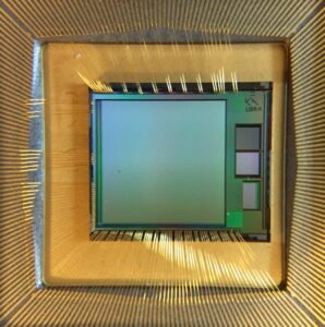
Dr. Scott will discuss a one-megapixel X-ray imaging detector with 7.8 µm pixel dimensions and high detection efficiency for hard X-ray energies above 20 keV. The amorphous selenium/CMOS detector technology reported can address gaps in commercially available X-ray detectors that limit their usefulness for existing applications such as phase contrast tomography and high-resolution imaging of nanoscale lattice distortions in bulk crystalline materials using Bragg coherent diffraction imaging at energies higher than 50 keV. The technology will also facilitate the creation of novel beamline imaging applications at X-ray energies at or above 20 keV. A second version of KA Imaging’s BrillianSe has also been developed with a 16megapixel detector.

The annual joint users meeting of the Center for Nanoscale Materials (CNM) and the co-located Advanced Photon Source (APS) will be held virtually in 2021 by Argonne National Laboratory. Anyone is welcome to register for myriad activities including technical workshops that highlight, promote, and stimulate user science; plenary sessions; poster sessions; a vendor expo; and CNM short courses. https://www.anl.gov/cnm/apscnm-users-meetings 

About KA Imaging 

A spin-off from the University of Waterloo, KA Imaging specializes in developing innovative X-ray imaging technologies and systems, providing solutions to the medical, veterinary, and non-destructive test industrial markets. For more information, please visit www.kaimaging.com.  

Contacts: 

Fernanda Fraga  

Media Relations 
ffraga@kaimaging.com  
T: 226.215.9897

Sensor at the core of the new detector

,

KA Imaging Creates Subscription Model for Reveal™ 35C to Support Hospitals During Pandemic

Hospitals are facing financial pressures despite the increasing need for COVID-19 care because outpatient and non-emergency procedures are declining. The American Hospital Association estimates that American hospitals incurred more than $202 billion in losses between March 1 and June 30 of this year. “COVID-19 has identified a significant problem in the healthcare industry,” said Karim […]

inCiTe Photo Friday: Connections in Flexible Electronics

Flexible electronics is a technology making remarkable progress and increasing in importance in many sectors including healthcare, automotive and retail. It allows electronic circuits to be built on flexible substrates, which makes them bendable and stretchable. Flexible, thin and light electronics allow designers to keep creating new products in innovative ways, but because the electronics […]