
About admin
This author has not written his bio yet.
But we are proud to say that admin contributed 81 entries already.
Entries by admin
World Health Day 2022: Our Planet, Our Health
World Health Day is celebrated every year on April 7th. The idea is to engage people in specific health related topics. “Our planet, our health” is this year’s theme.
Healthcare is governed by metrics of quality, cost, and access. When technologies come out, they tend to focus on just one of those aspects.
Over the past couple of years, we have seen immense change in many ways. At KA Imaging, we believe in making a difference in healthcare, pushing the norms of common X-ray practices. With Reveal 35C’s dual-energy subtraction (DES) technology, we aim to provide every facility with high-quality insights on diseases by enhancing X-ray quality, reducing costs, and making better imaging accessible for everyone. By working to achieve innovative X-ray everywhere, we can positively impact lives.
This is our planet, our health.
KA Imaging at RSNA 2021: focus on portability, confidence and speed
Two words can define KA Imaging’s first in-person event since the start of the pandemic: busy and successful. “It was a great opportunity to meet the imaging and industry communities,” said Amol Karnick, President and CEO of KA Imaging.
RSNA 2021 Scientific Assembly & Annual Meeting took place in Chicago from November 28 to Dec 02. It is the world’s largest medical imaging conference and showcases the latest medical imaging technology in X-ray, CT, MRI, artificial intelligence (AI), 3D printing, and more.
At RSNA 2021, KA Imaging showcased its innovative Reveal 35C, the worlds first single exposure dual-energy X-ray detector. Reveal is retrofittable and can be called a detector-side dual-energy upgrade: simply by replacing the detector, any existing X-ray system can be upgraded to dual-energy. With the same radiation dose as a chest X-ray, it is possible to create 3 different images without motion artifacts (the standard digital X-ray, soft tissue and bone), giving the ability to see the lungs and soft tissue without having bones obstructing the view, as well as to identify calcified nodules and vessels.
Dual-Energy Subtraction (DES) has been clinically shown to improve the detection of several chest diseases1. In a paper presented earlier this year at the European Congress of Radiology (ECR), the Reveal 35C technology was shown to detect 25% more pneumonia cases (including COVID-19) than traditional X-Ray2. “ The use of dual-energy x-ray in COVID-19 to detect manifestations of the disease in the lungs may contribute to improve diagnosis,” explained Karim S. Karim, CTO of KA Imaging. “The healthcare community has been seeking solutions that can be easily deployed, accurate and quickly disinfected – exactly what we want to provide with Reveal 35C,” he continued.
The company also presented some of what the engineering team has been developing behind the lab walls: single exposure dual energy tomosynthesis (an innovation showcase only, not available for sale). The first images were released just before the show. The idea is to provide DE tomo images using the existing clinical tomo techniques, lowering the radiation dose and eliminating motion artifacts.
References:
(Lung Nodules) Oda, Seitaro, Kazuo Awai, Yoshinori Funama, Daisuke Utsunomiya, Yumi Yanaga, Koichi Kawanaka, Takeshi Nakaura et al. “Detection of small pulmonary nodules on chest radiographs: efficacy of dual-energy subtraction technique using flat-panel detector chest radiography.” Clinical radiology 65, no. 8 (2010): 609-615.
(Pneumothorax) Urbaneja, A., Dodin, G., Hoosu, G., et al. (2018) Added Value of Bone Subtraction in Dual-energy Digital Radiography in the Detection of Pneuomothorax: Impact of Reader Expertise and Medical Specialty. The Association of University Radiologists. Elsevier Inc.
(Pneumonia) Martini, Katharina, Marco Baessler, Stephan Baumueller, and Thomas Frauenfelder. “Diagnostic accuracy and added value of dual-energy subtraction radiography compared to standard conventional radiography using computed tomography as standard of reference.” PloS one 12, no. 3 (2017): e0174285.
(Tuberculosis) Sharma, Madhurima, Manavjit Singh Sandhu, Ujjwal Gorsi, Dheeraj Gupta, and Niranjan Khandelwal. “Role of digital tomosynthesis and dual energy subtraction digital radiography in detection of parenchymal lesions in active pulmonary tuberculosis.” European Journal of Radiology 84, no. 9 (2015): 1820-1827.
(Coronary Calcifications) Song, Yingnan, Hao Wu, Di Wen, Bo Zhu, Philipp Graner, Leslie Ciancibello, Haran Rajeswaran et al. “Detection of coronary calcifications with dual energy chest X-rays: clinical evaluation.” The International Journal of Cardiovascular Imaging (2020): 1-8.
Kuhlman, Janet E., Jannette Collins, Gregory N. Brooks, Donald R. Yandow, and Lynn S. Broderick. “Dual-energy subtraction chest radiography: what to look for beyond calcified nodules.” Radiographics 26, no. 1 (2006): 79-92.
- Sanchez F, Kandel S, May M, Ronghe S, Rogalla P. Diagnostic value of dual-energy chest x-ray in immunocompromised patients to rule out pneumonia: initial results. European Congress of Radiology-ECR 2021, 2021
Ka Imaging to Exhibit at Arab Health 2022
The Company Is Part of The Ontario Life Science Mission
KA Imaging is going to participate in the largest health care event in the MENA region – Arab Health. KA Imaging is part of the Ontario Pavilion (that has a total of 17 companies).
When And Where:
Date: 24-27 January, 2022
Ontario booth: # SA N50
Address: Dubai World Trade Centre – Sheikh Zayed Rd, Dubai United Arab Emirates
KA Imaging will be showcasing Reveal 35C which is the world’s first single exposure portable dual-energy X-ray detector. Reveal is retrofittable and can be called a detector-side dual-energy upgrade: simply by replacing the detector, any existing X-ray system can be upgraded to dual-energy. With the same radiation dose as a chest X-ray, it creates 3 images without motion artifacts (standard digital X-ray, soft tissue and bone), giving the ability to see the lungs and soft tissue without having bones obstructing the view.
Arab Health is the Middle East’s largest life sciences trade show. The exhibition attracts over 100,000 attendees and 4,000 exhibiting companies. For 45 years, Arab Health has featured the latest innovations in healthcare. From state-of-the-art imaging equipment to the most cost-effective disposables, developments in surgery to advances in prosthetics, Arab Health continues to be at the heart of healthcare in the Middle East.
To learn more: https://www.arabhealthonline.com/en/Home.html
About KA Imaging
A spin-off from the University of Waterloo, KA Imaging specializes in developing innovative X-ray imaging technologies and systems, providing solutions to the medical, veterinary, and non-destructive test industrial markets. For more information, please visit www.kaimaging.com.
A Collection of Interesting Facts About X-ray Through the Years
KA Imaging loves celebrating innovation and the history of X-ray technology. Here are some facts we found intriguing and fun:
1. The most powerful X-ray generator has an output 80 times that of all the world’s electric generators.
The Z Pulsed Power Facility, known as the Z Machine, made at the Sandia National Laboratories, USA, and refurbished in 1996, is used to study extreme conditions in physics, like the nature of black holes1. It is the largest high-frequency electromagnetic wave generator and is used to gather data for nuclear research and development.
2. The most powerful X-ray telescope has the resolving power equivalent to the ability to read a stop sign at 12 miles away.
The Chandra, made in July of 1999, orbits the earth 200 times higher than Hubble, more than 1/3 of the way to the moon2. This telescope, with four very sensitive mirrors, detects X-ray emissions from stars, galaxies and black holes3.
3. An X-ray image was essential for developing a model of human DNA.
Photo 51 was taken by Raymond Gosling, a student of Rosalind Franklin, in May 1952 at King’s College London. This X-ray diffraction image of a gel with DNA fiber was used for developing a model of human DNA structure4.
4. X-rays were used in shoe stores from the 1920s to the 1970s.
Shoe-fitting fluoroscopes were used in shoe stores around the world and were often used to measure the feet of young children. They looked like a wooden box or short column, with an opening for customers to place their feet. The X-ray view of their feet could be seen through portholes at the top of the box5.
5. More than a million wounded soldiers in WWI had X-ray exams due to the efforts of Madame Marie Curie.
Marie Curie brought radiological cars right to the battlefields of France with the help of the Union of Women and 150 female volunteers. Curie’s cars contained an X-ray machine and photographic darkroom equipment, and used an electric generator. Curie even learned to drive and repair her donated vehicles6.
6. X-rays can see old paintings
It is absolutely fascinating that x-ray technology can be used more than just in the medical world. When art historians want to know if there was something painted on a canvas before the final work of art, they can use x-rays to determine that7.
7. X-ray named top achievement by British museum
X-Ray was named the most important modern scientific discovery exceeding 50K votes in a Science Museum of London poll, even surpassing penicillin8!
References
1. Most powerful X-ray generator.
2. Most powerful X-ray telescope.
4. Photo 51.
6. Marie Curie.
7. Vermeer, Girl with a Pearl Earring .
8. British museum.
KA Imaging’s Reveal 35C Shown to Find More Pneumonia Cases than Traditional X-Ray
Company will be exhibiting at 2021 RSNA Scientific Assembly and Annual Meeting
Waterloo, Ont. (Nov 17, 2021)
“Data drives decisions and with the latest trial data, we are positioned for growth,” says Amol Karnick, President and CEO of KA Imaging. In a paper presented earlier this year, the Reveal 35C technology was shown to detect 33% more pneumonia cases (including COVID-19) than traditional X-Ray1.
Reveal 35C is the world’s first single exposure portable dual-energy X-ray detector. Reveal is retrofittable and can be called a detector-side dual-energy upgrade: simply by replacing the detector, any existing X-ray system can be upgraded to dual-energy. With the same radiation dose as a chest X-ray, it creates 3 images without motion artifacts (standard digital X-ray, soft tissue and bone), giving the ability to see the lungs and soft tissue without having bones obstructing the view.
The paper presents the initial results of an ongoing clinical trial that is being carried out in Toronto (Canada). “Although Reveal has been available for sale in both the US and Canada for the past year, we have enrolled Reveal in a number of clinical trials to demonstrate the significant improvement in clinical outcomes compared to standard digital X-ray,” says Karnick.
According to the company, Reveal 35C enhances the use of dual energy, benefiting not only radiologists, but the entire service chain.
“It’s good for radiologists, who can access supplementary dual energy X-ray images for faster and easier reading,” explains Karnick. “It’s good for imaging technologists, who can offer patients more comfort by bringing the detector to the point of care and getting high-quality images even in challenging settings such as critical care units; it’s good for administrators, who can optimize the use of other equipment across multiple departments; and it is especially good for patients, who have more comfort and early access to treatment for diseases that could go unnoticed until they become more serious”, he concludes.
KA Imaging at RSNA
KA Imaging is exhibiting at 2021 RSNA Scientific Assembly and Annual Meeting. The company is located at booth 6544 in McCormick Place’s North Hall level 3.
- Sanchez F, Kandel S, May M, Ronghe S, Rogalla P. Diagnostic value of dual-energy chest x-ray in immunocompromised patients to rule out pneumonia: initial results. European Congress of Radiology-ECR 2021, 2021.
KA Imaging Adds Ted Sloan to Its Executive Team
Manufacturer KA Imaging has appointed Ted Sloan to its executive team. Ted will be leading the company’s channel strategy, strengthening its business development efforts.
For over 30 years, Ted Sloan held several executive roles at Merry X-ray Corporation/MXR Imaging, leading the company as President from 2012 to 2021. Ted Sloan’s business acumen is rooted in ground-up sales experience and technical training, which will make him a major contributor to KA’s growth.
Earlier this year, the company named other healthcare veterans to key leadership roles: Shawn Campbell as Vice President of Operations and Robert Moccia as Vice President of Sales, USA and Canada.
World Radiology Day
Happy World Radiology Day! Every year on Nov 8th, the world commemorates the discovery of X-rays in 1895. World Radiology Day is observed to improve public awareness of radiographic imaging and therapy, which are critical in patient diagnosis and treatment.
Find out more about the advantages of x-rays.
REFERENCES
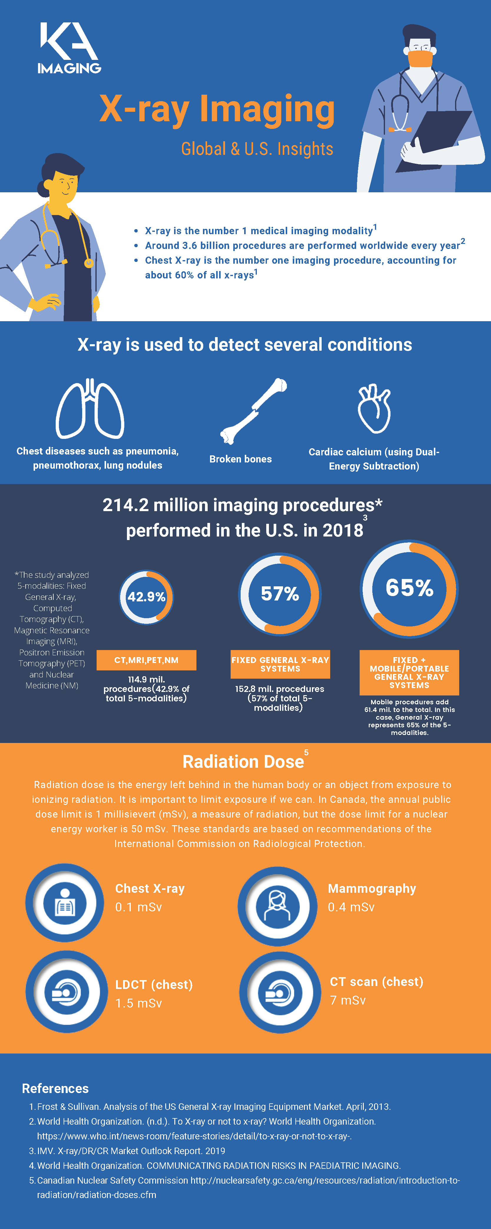
- Frost & Sullivan. Analysis of the US General X-ray Imaging Equipment Market. April, 2013.
- World Health Organization. (n.d.). To X-ray or not to x-ray? World Health Organization. https://www.who.int/news-room/feature-stories/detail/to-x-ray-or-not-to-x-ray-.
- IMV. X-ray/DR/CR Market Outlook Report. 2019
- World Health Organization. COMMUNICATING RADIATION RISKS IN PAEDIATRIC IMAGING.
- Canadian Nuclear Safety Commission http://nuclearsafety.gc.ca/eng/resources/radiation/introduction-to-radiation/radiation-doses.cfm
KA Imaging at RSNA 2021
KA Imaging is ready to take on the biggest event of the year. Visit us at RSNA 2021, North Hall Level 3, Booth 6544. RSNA 2021 is the world’s largest medical imaging conference and showcases the latest medical imaging technology in X-ray, CT, MRI, artificial intelligence (AI), 3D printing, and more. We’re looking forward to meeting you!
Going to RSNA 2021 in Chicago? Make time in your schedule to visit KA Imaging’s booth in the North Hall. Get informed. Get energized! See innovation in action. Send us an email to schedule your appointment with one of our representatives today!
When and Where
Technical Exhibition Date: November 28 – December 1
Booth Location: RSNA 2021, North Hall Level 3, Booth 6544.
Address: McCormick Place
2301 S King Drive, Chicago, IL 60616
What to expect
KA Imaging will be exhibiting a few of the products currently in the roster at RSNA.
Reveal™ 35C
Reveal™ can replace existing x-ray detectors and provide radiologists with unobstructed front and lateral views of the lungs and bones. Reveal is the world’s first portable dual-energy detector, and can be taken to the bedside of patients, as well as in the field.
InCiTe™ 3D X-ray Microscope
The inCiTe™ 3D X-ray Microscope provides phase contrast imaging and micro-CT capabilities from KA Imaging, and is the first commercial X-ray CT system that utilizes BrillianSe™.
Sight™
Sight™ is a flat panel detector which features a light-weight design and low X-ray dose. Improved Modulation Transfer Function (MTF) and Detective Quantum Efficiency (DQE) provide uncompromising image quality.
3D Virtual demo
KA Imaging is finalizing the last few pieces in the puzzle to gear up a 3D Virtual Demo of Reveal 35C, stay tuned as more information will be shared shortly!
November Webinar Events
November will be a busy month for KA Imaging, with 3 webinars already scheduled, each tailored for a specific audience.
On November 16, Radiology Managers and Imaging Directors will have the opportunity to attend the webinar “Improving Department Results with Single Exposure Dual Energy Subtraction X-Ray: Clinical, Operational and Financial Benefits”. This webinar is partnered with ICE Magazine and is pending approval by the AHRA for 1.0 Category A ARRT. Dr. Karim will be available for a live Q&A session!
On November 18, Dr. Karim S. Karim and radiologist Dr. Patrik Rogalla will present the Reveal 35C to the Canadian Pulmonary Fibrosis Foundation. Dr. Patrik will show you some of the stunning images captured in a clinical trial he is conducting at a hospital in Toronto.
On November 20th, it will be the imaging technicians’ turn to learn more about dual energy. The “Dual Energy Radiography” webinar is specially designed for these professionals and is approved by the Canadian Association of Medical Radiation Technologists (CAMRT) for up to 1.75 credit hours of Category a Continuing Education.
Visit the links below to learn more and register!
Nov 16 – 2:00 PM EST
Certification: Pending approval by AHRA for 1.0 Category A ARRT
Register here: https://register.gotowebinar.com/register/8154594376304538124
Nov 18 – 1:30 PM EST
Speakers: Dr. Karim Karim & Dr. Patrik Rogalla
Register here: https://www.hopebreatheshere.com/live-webinars
Nov 20 – 10:00 AM EST
Speakers: Dr. Karim Karim & Jay Potipcoe
Certification: Approved by the Canadian Association of Medical Radiation Technologists (CAMRT) for up to 1.75 hours Category A credit in continuing Education; recognized by the CAMRT’s provincial organizations and the American Registry of Radiologic Technologists (ARRT).
Register here: https://vcaeducation.ca/event/dual-energy-radiography-live-webinar-nov202021/
Media Inquiries