

The flexibility
of a 3-in-1
solution
What if you could improve image quality and get bone and tissue differentiation from your fixed or mobile system, keeping the ease and simplicity of X-ray? With Reveal™ 35C, You can.
Reveal™ 35C is the first and only mobile dual-energy detector on the market. Thanks to its SpectralDR™ technology, it offers improved visualization of lung nodules, pneumonia, line and tube tips, pneumothorax, retained surgical objects and more. This 3-in-1 solution simultaneously acquires 3 images in 1 single exposure, improving visualization of bone and tissue.

Drag the slider to transition between a traditional DR and the SpectralDR™ images created by Reveal.
In this real study case involving a 51-year-old female leukemia patient, the Conventional DR X-Ray was read as normal. Upon viewing the Dual-Energy Soft Tissue image, the radiologist noticed a highlighted focal opacity indicative of pneumonia (confirmed on CT).
![]()
1 exposure, 3 different images, 0 motion artifacts
![]()
33% more pneumonia cases found compared to x-ray thanks to dual-energy images4
![]()
Same dose. Same source. Same technique.
![]()
45% more lesion visibility when SpectralDR™ images were included5.
![]()
No disruption to workflow.
![]()
DQE as high as 75%
![]()
The only mobile dual-energy solution
![]()
INCREASE CONFIDENCE. Improve your diagnosis.
Why choose SpectralDR™ technology? Dual energy technology has the potential to change the world of X-ray for the better. However, it is known for having numerous limitations in multiple fields. Our SpectralDRTM technology addresses each of those limitations to ensure the satisfaction and confidence for every user involved.
Contact us to REQUEST A FREE DEMO
Better financial outcomes
In addition to the clinical benefits, Reveal 35C can help imaging departments to improve their financial outcomes. Visit our blog to learn about the high potential economic impact of Reveal 35C.
Reveal 35C advantages
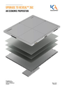
Reveal 35C uses SpectralDR™ technology to acquire three images simultaneously (DR, bone and soft tissue dual energy X-ray images) with only a single standard X-ray exposure. The technology mimics the workflow, dose and techniques of state-of-the-art mobile DR digital X-ray detectors.
Reveal can replace existing x-ray detectors and provide radiologists with unobstructed front and lateral views of the lungs and bones, which can aid in the visualization of pneumonia, fractures, catheters (i.e. tubes and PICC lines), coronary calcium, and masses with high sensitivity. The soft tissue and bone images are sharp and free of motion artifacts, which increases the diagnostic sensitivity. Reveal 35C can be taken to the bedside of patients, as well as in the field.

Improved patient outcomes
Early disease detection shortens time to providing corrective procedures1

Enhanced patient and operator safety
20X less radiation compared to CT2
Reduction in diagnostic errors and malpractice concerns2

Flexible applications
Portability allows the detector to be taken where it is most needed.

Universally compatible
ISO Cassette size
Use your existing infrastructure
Higher operating efficiencies
Reduces radiologist reading time for X-rays by 30%3
Enables residents to make accurate diagnoses3

Significant savings
10X lower purchase and operating costs than CT
Reveal™ 35C is available in Canada, the USA, Australia, New Zealand, Taiwan, Indonesia and Malaysia. Contact us for availability in other countries.
References
1. Improved patient outcomes
(Lung Nodules) Oda, Seitaro, Kazuo Awai, Yoshinori Funama, Daisuke Utsunomiya, Yumi Yanaga, Koichi Kawanaka, Takeshi Nakaura et al. “Detection of small pulmonary nodules on chest radiographs: efficacy of dual-energy subtraction technique using flat-panel detector chest radiography.” Clinical radiology 65, no. 8 (2010): 609-615.
(Pneumothorax) Urbaneja, A., Dodin, G., Hoosu, G., et al. (2018) Added Value of Bone Subtraction in Dual-energy Digital Radiography in the Detection of Pneuomothorax: Impact of Reader Expertise and Medical Specialty. The Association of University Radiologists. Elsevier Inc.
(Pneumonia) Martini, Katharina, Marco Baessler, Stephan Baumueller, and Thomas Frauenfelder. “Diagnostic accuracy and added value of dual-energy subtraction radiography compared to standard conventional radiography using computed tomography as standard of reference.” PloS one 12, no. 3 (2017): e0174285.
(Tuberculosis) Sharma, Madhurima, Manavjit Singh Sandhu, Ujjwal Gorsi, Dheeraj Gupta, and Niranjan Khandelwal. “Role of digital tomosynthesis and dual energy subtraction digital radiography in detection of parenchymal lesions in active pulmonary tuberculosis.” European Journal of Radiology 84, no. 9 (2015): 1820-1827.
(Coronary Calcifications) Song, Yingnan, Hao Wu, Di Wen, Bo Zhu, Philipp Graner, Leslie Ciancibello, Haran Rajeswaran et al. “Detection of coronary calcifications with dual energy chest X-rays: clinical evaluation.” The International Journal of Cardiovascular Imaging (2020): 1-8.
Kuhlman, Janet E., Jannette Collins, Gregory N. Brooks, Donald R. Yandow, and Lynn S. Broderick. “Dual-energy subtraction chest radiography: what to look for beyond calcified nodules.” Radiographics 26, no. 1 (2006): 79-92.
2. Enhanced patient and operator safety
Kuhlman, Janet E., Jannette Collins, Gregory N. Brooks, Donald R. Yandow, and Lynn S. Broderick. “Dual-energy subtraction chest radiography: what to look for beyond calcified nodules.” Radiographics 26, no. 1 (2006): 79-92.
3. Higher operating efficiencies
Manji, Farheen, Jiheng Wang, Geoff Norman, Zhou Wang, and David Koff. “Comparison of dual energy subtraction chest radiography and traditional chest X-rays in the detection of pulmonary nodules.” Quantitative imaging in medicine and surgery 6, no. 1 (2016): 1.
4. 33% more pneumonia cases
Sanchez F, Kandel S, May M, Ronghe S, Rogalla P. Diagnostic value of dual-energy chest x-ray in immunocompromised patients to rule out pneumonia: initial results. European Congress of Radiology-ECR 2021, 2021
5. 45% more lesion visibility
S. L. Maurino, K. S. Karim, V. Venkatesh. Diagnostic value of single‐exposure dual‐energy subtraction
radiography in lung lesion detection: initial results. European Congress of Radiology-ECR
2022, 2022
Media Inquiries