
inCiTe™ 3D X-ray Microscope
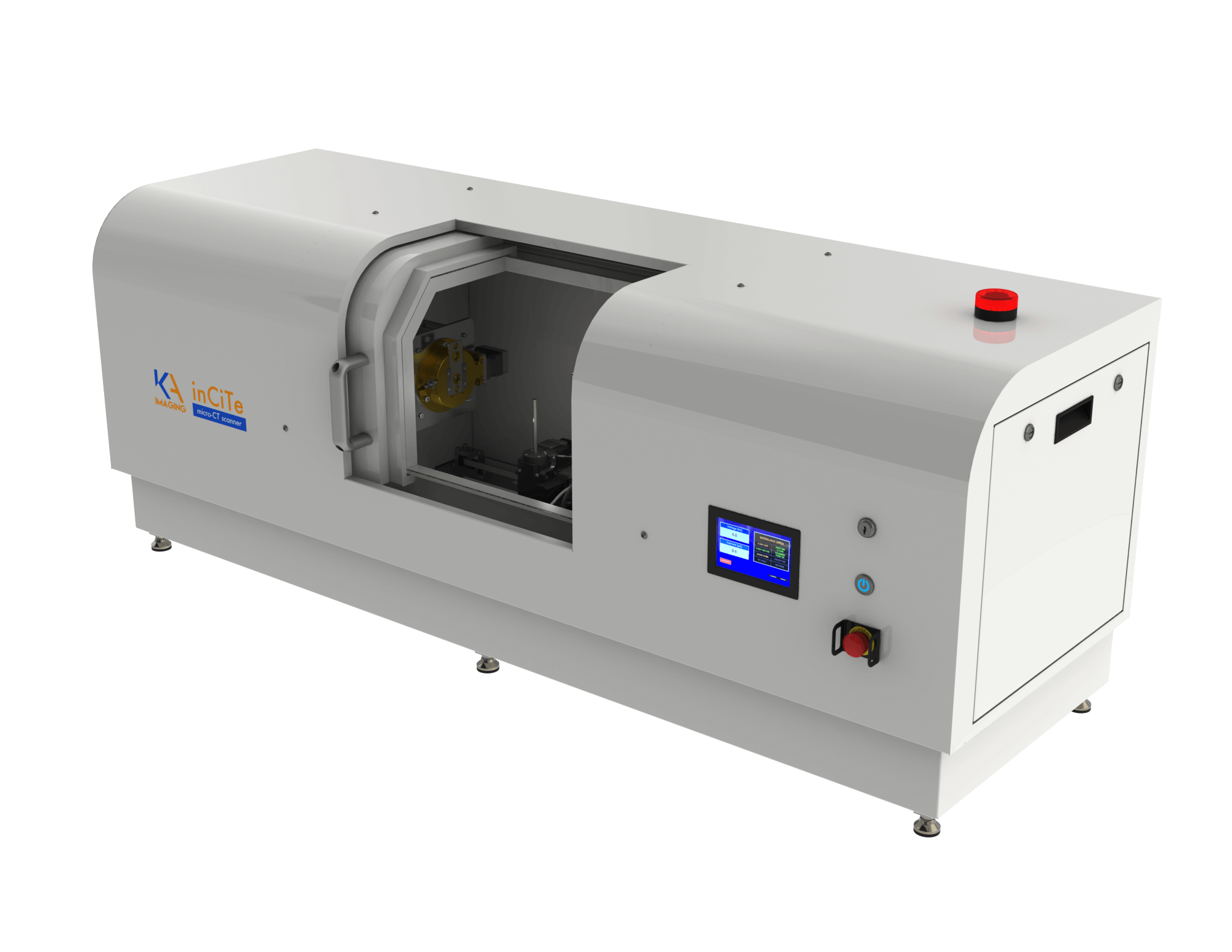
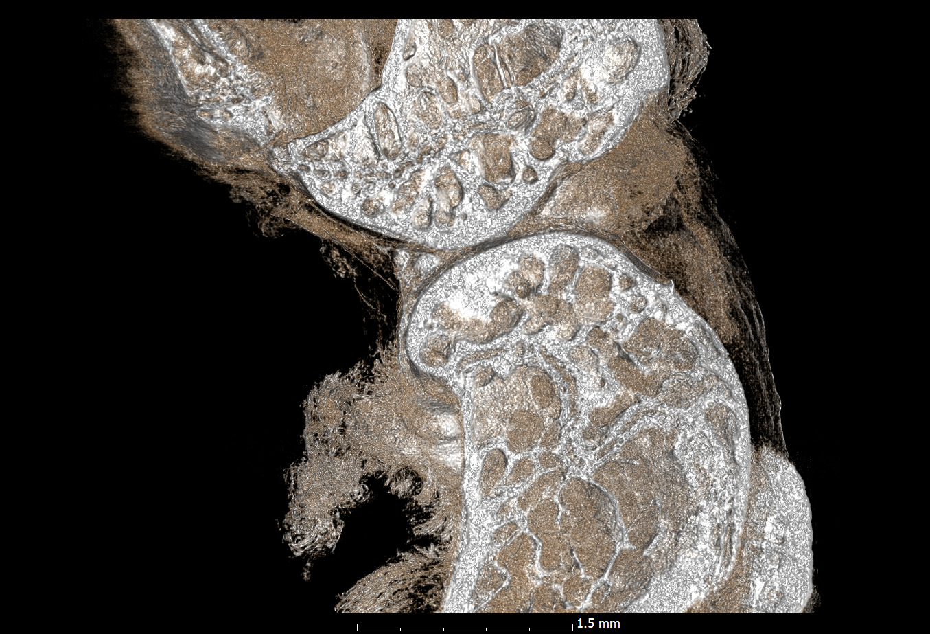
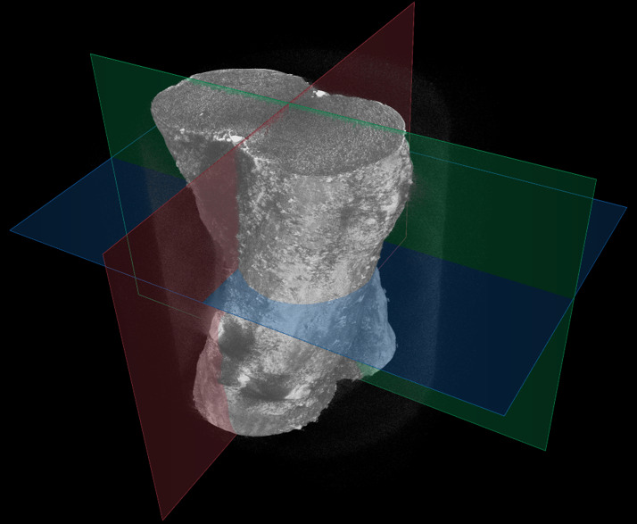
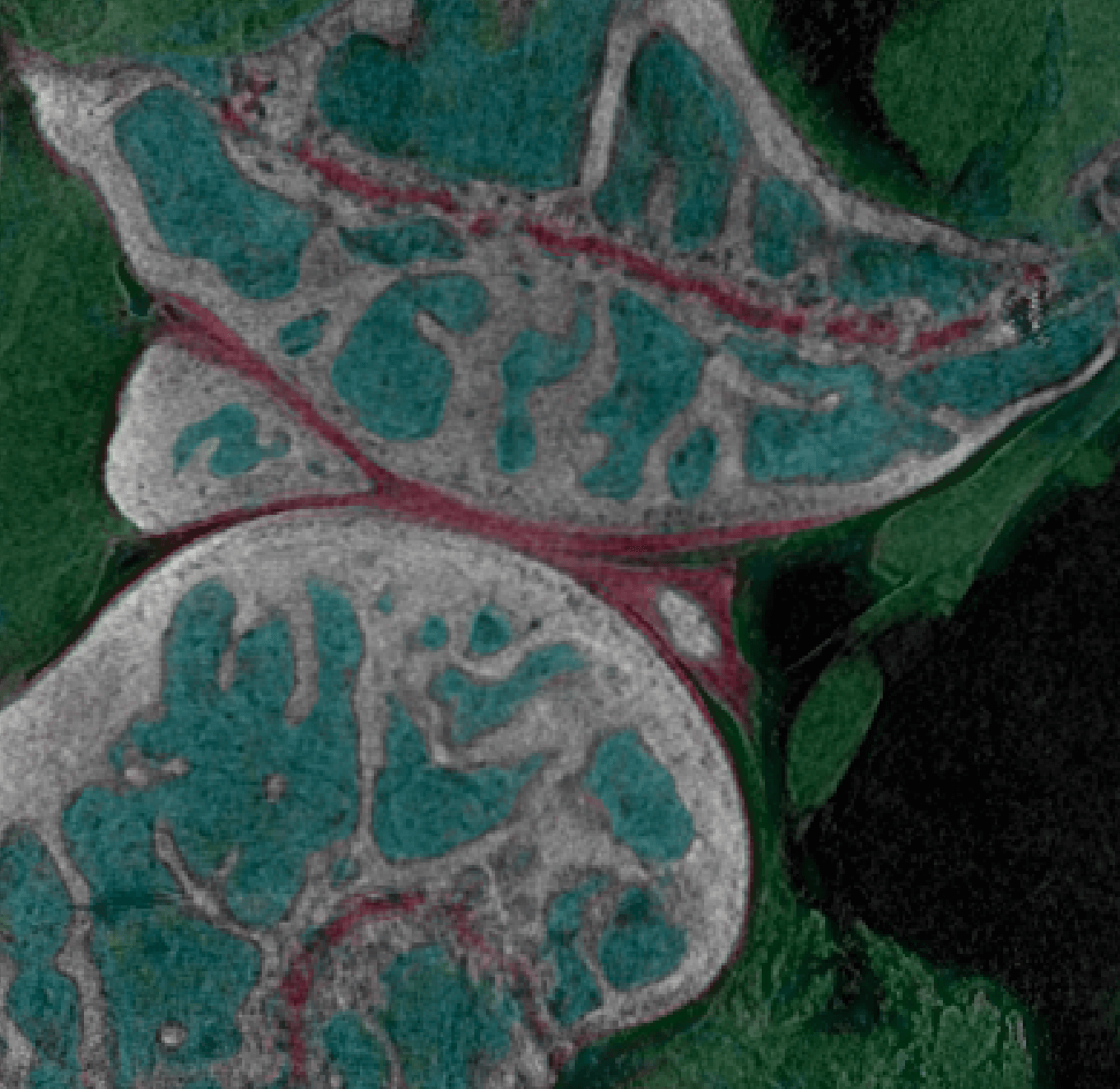
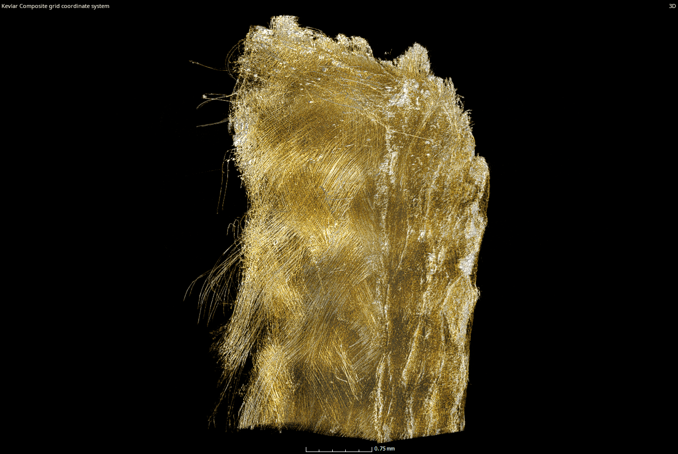
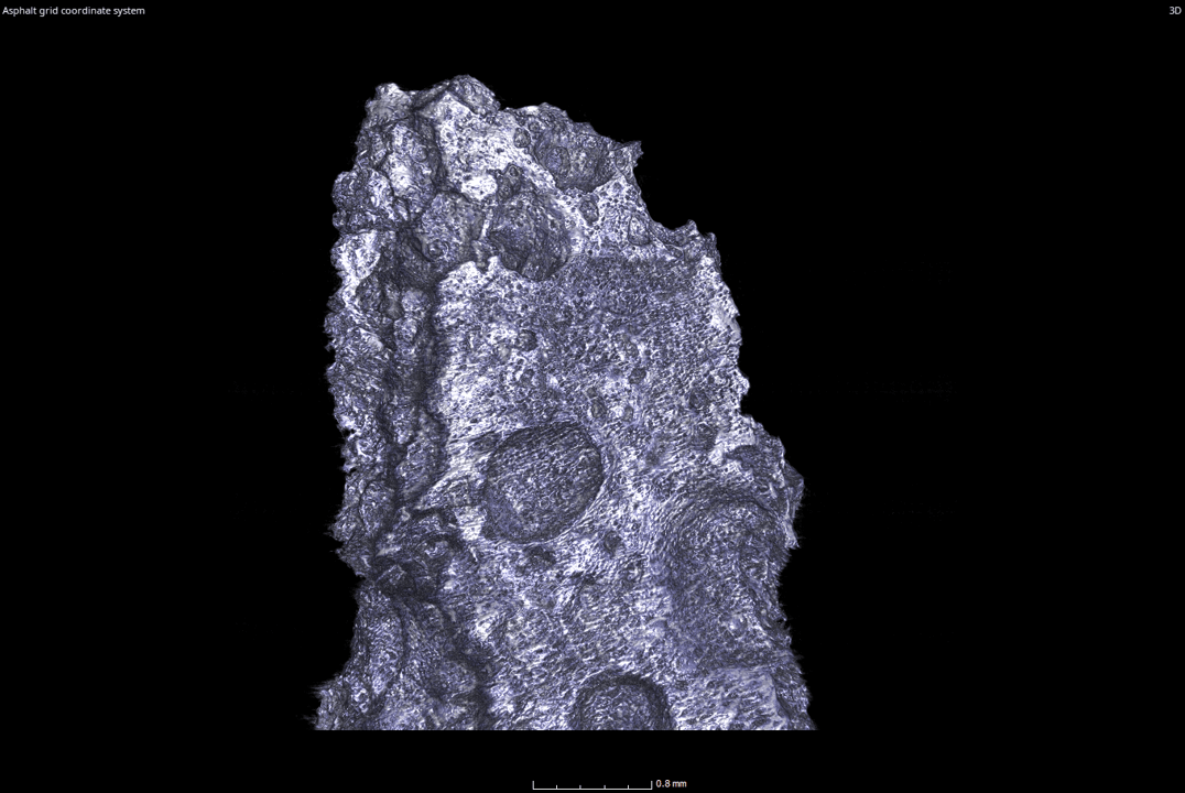
The inCiTe™ 3D X-ray Microscope is a state-of-the-art benchtop Micro-CT system designed for unparalleled imaging clarity, especially in low-density materials. With advanced phase contrast capabilities, inCiTe™ reveals intricate details in soft biological tissues, polymers, and other challenging samples that traditional X-ray methods struggle to visualize. Compact yet powerful, it provides researchers and professionals with high-resolution, three-dimensional insights, revolutionizing applications in material science, biomedical research, and quality control.
inCiTe™ is the first commercial X-ray CT system to utilize the BrillianSe™ X-ray Detector. BrillianSe™, our patented amorphous selenium (a-Se) detector, offers a unique combination of high spatial resolution and detection efficiency
Product overview
X-ray detector: 16MP CMOS Detector, 8μm pitch, 14 bit resolution
Spatial resolution: 11 μm ( 0.5 MTF at 45 cycles/mm); 5.6 μm ( 0.1 MTF at 90 cycles/mm
X-ray Source: 40-110 KV, 16 W, 2 μm spot size
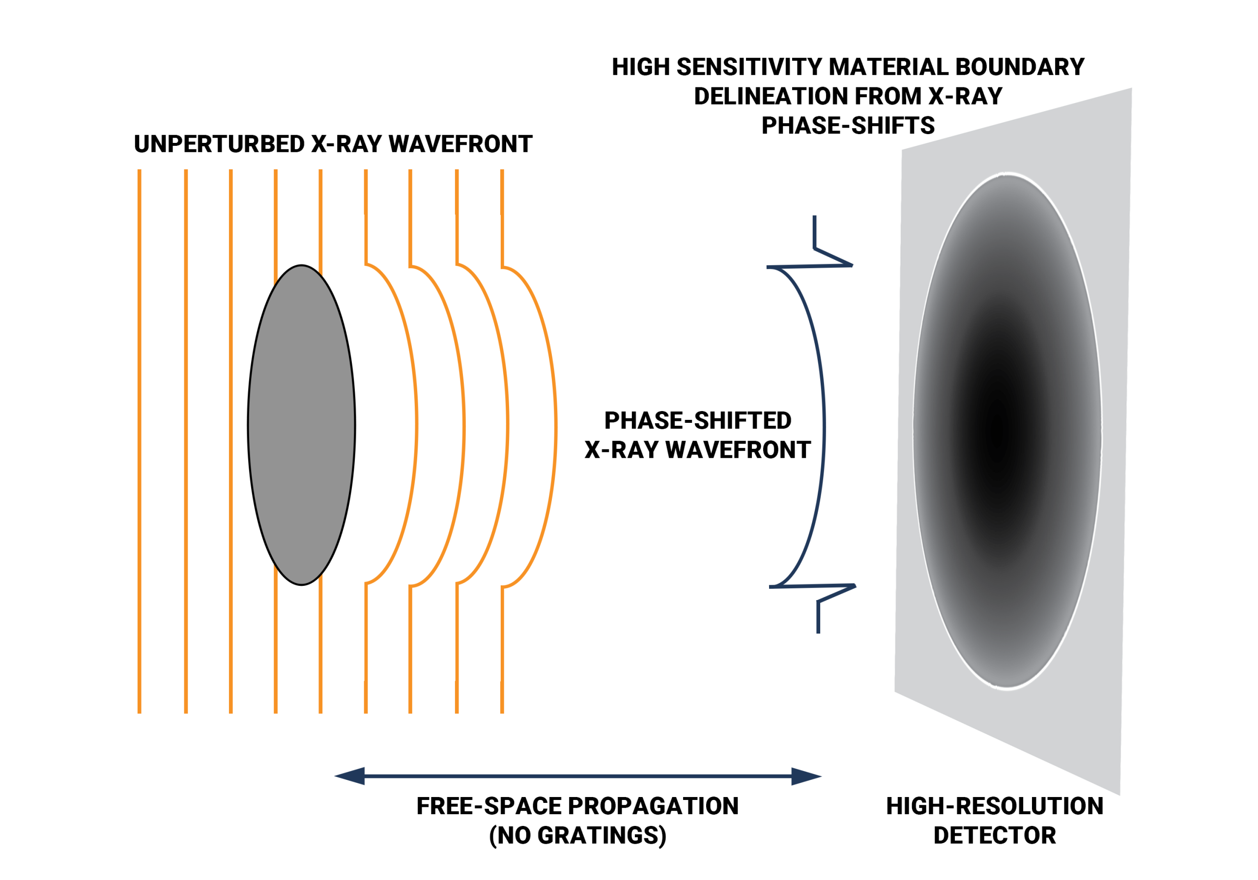
Propagation-based X-ray phase contrast
Suitable for synchrotron applications
Faster scan time
Literature
inCiTe™: Advancing 3D X-ray Imaging and Non-Destructive Testing (NDT)
inCiTe™, our cutting-edge 3D X-ray microscope, sets a new standard in scientific imaging and non-destructive testing (NDT). Powered by the innovative BrillianSe™ X-ray Detector, inCiTe™ delivers exceptional clarity and precision. It revolutionizes scientific research and NDT in various applications:
Non-destructive Testing (NDT)
Additive manufacturing
Electronics
Agriculture
Geology
Preclinical imaging
Customer Stories
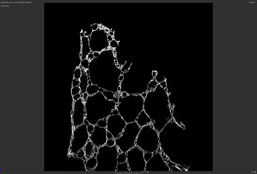
Phase Contrast: better visualization for low-density materials
The inCiTe™ 3D X-ray Microscope excels in phase-contrast imaging, which is crucial when traditional X-ray methods struggle with low-density materials like soft tissues or polymers. Unlike conventional systems that measure only X-ray intensity, thanks to BrillianSe™ X-ray Detector, inCiTe™ can capture subtle phase shifts, enhancing image contrast. This is achieved using free-space propagation, where phase variations translate directly into intensity fluctuations, offering clearer, more detailed images.
About BrillianSe™ X-ray Detector
BrillianSe™ is a hybrid a-Se/CMOS detector that uses an a-Se photoconductor with high intrinsic spatial resolution for direct conversion of X-ray photons to electric charge. The electronic signal is then read out by a low noise CMOS active pixel sensor (APS). Without the need to first convert X-ray photons to visible light, as in indirect scintillator-based approaches, thinning of the conversion layer to minimize optical scatter is not necessary.
Latest blogs
KA Imaging’s patented amorphous selenium (a-Se) BrillianSe™ X-ray imaging detectors have been designed into the…
In the realm of industrial safety and quality assurance, Non-destructive Testing (NDT) emerges as a…
Original research
S. Abbaszadeh, CC Scott, O Bubon, A Reznik, KS Karim, “Enhanced detection efficiency of direct conversion X-ray detector using polyimide as hole-blocking layer,” Scientific Reports, December 2013
G.P. Lindberg, T. O’Loughlin, N. Gross, A. Mishchenko, A. Reznik, S. Abbaszadeh, K.S. Karim, G. Belev, B. Weinstein, “Photo-crystallization in a-Se layer structures: effects of film-substrate interface-rigidity,” Journal of Applied Physics, vol. 116, November 2014.
A. Parsafar, C. Scott, A. El-Falou, P. Levine, K.S. Karim, “Direct-Conversion CMOS X-Ray Imager with 5.6 μm × 6.25 μm Pixels,” IEEE Electron Device Letters, 36(5), pp. 481-3, May 2015.
C.C. Scott, A. Parsafar, A. El-Falou, P. M. Levine, K.S. Karim, “High Dose Efficiency, Ultra-high Resolution Amorphous Selenium/CMOS Hybrid Digital X-ray Imager,” IEEE International Electron Devices Meeting (IEDM) Technical Digest, December 2015.
Related research
- Comparing conventional and phase contrast imaging
Gureyev, Timur Eugenievich, Sheridan C. Mayo, Damian E. Myers, Ya Nesterets, D. M. Paganin, A. Pogany, Andrew W. Stevenson, and S. W. Wilkins. “Refracting Röntgen’s rays: propagation-based x-ray phase contrast for biomedical imaging.” Journal of Applied Physics 105, no. 10 (2009): 102005.
Krenkel, Martin, Mareike Töpperwien, Christian Dullin, Frauke Alves, and Tim Salditt. “Propagation-based phase-contrast tomography for high-resolution lung imaging with laboratory sources.” AIP Advances 6, no. 3 (2016): 035007.
Olivo, A., and E. Castelli. “X-ray phase contrast imaging: From synchrotrons to conventional sources.” La Rivista Del Nuovo Cimento 37 (2014): 467-508.
Bravin, Alberto, Paola Coan, and Pekka Suortti. “X-ray phase-contrast imaging: from pre-clinical applications towards clinics.” Physics in Medicine & Biology 58, no. 1 (2012): R1.
Bidola, P., K. Morgan, M. Willner, A. Fehringer, S. Allner, F. Prade, F. Pfeiffer, and K. Achterhold. “Application of sensitive, high‐resolution imaging at a commercial lab‐based X‐ray micro‐CT system using propagation‐based phase retrieval.” Journal of Microscopy 266, no. 2 (2017): 211-220.
- Propagation
Wilkins, S. W., T. Ei Gureyev, D. Gao, A. Pogany, and A. W. Stevenson. “Phase-contrast imaging using polychromatic hard X-rays.” Nature 384, no. 6607 (1996): 335-338.
Mayo, Sheridan C., Andrew W. Stevenson, and Stephen W. Wilkins. “In-line phase-contrast X-ray imaging and tomography for materials science.” Materials 5, no. 5 (2012): 937-965.
- Gratings
Pfeiffer, Franz, Timm Weitkamp, Oliver Bunk, and Christian David. “Phase retrieval and differential phase-contrast imaging with low-brilliance X-ray sources.” Nature physics 2, no. 4 (2006): 258-261.
